Download
1 / 28
280 likes | 304 Vues
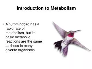
Explore exergonic and endergonic reactions, energy coupling, ATP, enzymes, enzyme inhibitors, and more in biochemical pathways. Learn about factors affecting enzyme activity and how inhibitors help control metabolic pathways.

E N D
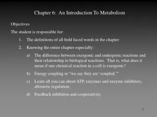
Chapter 6: An Introduction To Metabolism • Objectives • The student is responsible for: • The definitions of all bold faced words in the chapter • Knowing the entire chapter especially: • The difference between exergonic and endergonic reactions and their relationship to biological reactions. That is, what does it mean if one chemical reaction in a cell is exergonic? • Energy coupling or “we say they are ‘coupled.’” • Learn all you can about ATP, enzymes and enzyme inhibitors, allosteric regulation, • Feedback inhibition and cooperativity.
Figure 6.6 Energy changes in exergonic and endergonic reactions
Metabolic Disequilibium • What could a cellular reaction do when it reached equilibrium? • Well, it makes products at the same rate that the products get consumed and re-make the reactants. • Does this sound like the cell is carrying on any processes? • So, Le Chatelier’s principle tells us we can disturb the equilibrium to get a cell to do what we want. And what do we want the cell to do? Well generally we want the cell to carry out biochemical reactions. • So how do we get the biochemical reaction to “go forward?” Biochemical reactions are coupled.
Figure 6.9 Energy coupling by phosphate transfer Endergonic so it is not favored
The 3 phosphate groups are all negatively charged and make for weak bonds between the groups so they can be easily hydrolyzed. Figure 6.8 The structure and hydrolysis of ATP
Figure 6.8x ATP ATP ADP + Pi + 7.3 kcal at standard conditions but cells are not at standard conditions. In a cell the free energy change is – 13 kcal/ mole No such thing as a high energy bond But doesn’t the release of 13 kcal heat up the cell’s environment? No, because the energy is used to drive other reactions by phosphorylating other molecules
Figure 6.10 The ATP cycle The regeneration of ATP occurs at the rate of 10 million molecules of ATP per second in 1 cell.
Figure 6.11 Example of an enzyme-catalyzed reaction: Hydrolysis of sucrose Sucrase breaks down sucrose Why does sucrose taste sweeter than glucose?
Molecular Biology of the Cell Disk • Animation 2.1: Enzyme Catalysis
The active site is not a rigid area. Weak bonds are made for temporary bonding Enzyme can change its shape for better fit with substrate at the active site. This is the concept of “Induced Fit.” Figure 6.14 The induced fit between an enzyme and its substrate
Rate of Enzyme Activity and Substrate Concentration • Rate depends upon the concentration of the substrate. Does this make sense? • If you add more substrate, more substrate can be broken down so the rate of the enzyme activity is higher. • Up to a point. . . What if you saturate the enzyme? Enzyme is saturated Rate Of Rxn Concentration of Substrate
Two fundamental factors affecting the nature of enzymes are (1) pH and (2) temperature. Enzymes become denatured and lose their function Many cofactors are needed: Zn2+, Fe2+, Cu2+, Se2+, Mg2+, vitamins Organic cofactors are called coenzymes. Figure 6.16 Environmental factors affecting enzyme activity Stomach Small Intestine
Competitive Inhibitors can be identified or distinguished by: • increasing the presence or concentration of the substrate which increases the effectiveness of the enzyme • the fact that they occupy the same site as the substrate • penicillin is a competitive inhibitor of an enzyme involved with bacterial cell wall synthesis • Noncompetitive Inhibitors: • bind to sites away from the active site on the enzyme and alter the conformation of the active site • cannot be overcome by increasing the concentration of substrate Inhibitors help to control metabolic pathways. . This is a good thing.
Allosteric Enzyme Regulation • Most enzymes demonstrate this kind of regulation • An allosteric regulator has: • a binding site for the substrate • a binding or regulatory site for the activator • a binding or regulatory site for an inhibitor A B C D E F E1 E2 E3 E4 E5 ( - ) (+) E1 could have 12 distinct regulatory sites.
Made up of several polypeptide chains and each subunit has its own active site so there can be several allosteric sites on the enzyme • Binding of regulator, positive or negative, is reversible. • Case 1 • Protein complex(active) Protein complex(inactive) • Cooperativity: The binding of one substrate to one site shifts the equilibrium to the left so the enzyme remains in the active form. This increases enzyme activity. So by adding more substrate you encourage the formation of the active form.
Rate of Enzyme Activity and Substrate Concentration: Again So with a little substrate, you stabilize a little of the allosteric enzyme and shift equilibrium to left. Then you have more and more of the stable, active form so rate of reaction increases Rate Of Rxn Enzyme is saturated Concentration of Substrate
Did Someone just whisper, “I wish he’d give us an example”? • Phosphofructokinase or PFK • Catalyzes the phosphorylation of fructose-6-phosphate to fructose 1,6- diphosphate • F-6-P + ATP F-1,6-DP + ADP • ATP is a negative effector or an allosteric inhibitor so ATP keeps PFK in its inactive state. • If you add ADP, this speeds up the reaction so ADP has a binding site to shift the equilibrium to the active state. • So PFK has: • An active site for F-6-P • A neg. regulatory site for ATP • A pos. regulatory site for ADP
Lesch-Nyhan Syndrome Biochemical Defect Enzyme called hypoxanthan-guanine phosphoribosyltransferase (HGPRT) If you lack this enzyme you want to mutilate yourself by biting your hands, lips, etc. It is asymmetric so you only bit one side. The affected individual is bandaged to prevent their behavior Other effects: retarded, aggressive, and attack their care givers.
Testicular Feminization Syndrome TF is when you are phenotypically a female but you have male hormonal levels so you can’t have kids, etc. and testosterone levels are very high. So if testosterone levels are high, why aren’t you a male? How is this disorder discovered?
Adenosine Deaminase (ADA) and the Boy in the Bubble ADA removes –NH2 groups from adenine One of the causes of Severe Combined Immunodeficiency or SCID when lymphocytes fail to develop normally. Without the enzyme ADA, deoxyadenosine accumulates which causes the build up of deoxyadenosine triphosphate which inhibits an enzyme ribonucleotide reductase. Ribonucleotide reductase helps to make 4 nucleotides needed for DNA synthesis. All cells have ADA but lymphocytes have only ADA to degrade deoxyadenosine while other cells have other enzymes to degrade it. So it is the lymphocytes that fail and the immune system falters miserably.
Therapies for ADA Deficiency • Bone Marrow Transplant with cells from sibling • Enzyme Replacement Therapy: functional ADA is injected by IV but this is not a cure • Gene Therapy: • take lymphocytes from the patient and grow them in culture. • Obtain a virus that has a functional ADA gene and let viruses infect the cultured lymphocytes. • return lymphocytes to patient (altered somatic cells not germ cells) • repeat many times (11-12) over a 2 year period • so you have an increase in the number of lymphocytes and an increase in immune function