Download
1 / 22
220 likes | 258 Vues
Explore the detailed functional anatomy of the pelvic region, including the genital organs, pelvis major and minor, pelvic girdle, birth canal, and more. This resource offers essential information and advice on learning anatomy effectively.

E N D
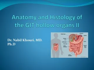
Ole Petter Ottersen Anatomy of the genital organs Jon Storm-Mathisen Department of Anatomy, Institute of Basic Medical Sciences & Centre for Molecular Biology and Neuroscience, University of Oslo, Norway jon.storm-mathisen@medisin.uio.no http://www.cmbn.no/group-storm-mathisen.html Per Brodal Undervisningsleder i anatomi
The Pelvic Region: Introduction to its Functional Anatomy Per Brodal Institutt for medisinske basalfag Universitetet i Oslo 9th Semester 2008
Anatomy 9th Semester • Lectures • gross anatomy, histology (microscopic anatomy), embryology • Intention: overviews, help to self-study • Practicals Histology • assumes a certain level of theoretical knowledge (lecture given a few days in advance of the practical classes) • Gross Anatomy Demos • three PBL-groups (20-25 students)
Demos, cont’d • Keep the group-size optimal: Meet at your scheduled time! • You are not allowed to switch to another group for convinience • Study the booklet for the Demos in advance (”Kompendium”, sold at the Akademika)
Assessment • Practical anatomy test (see page 41 in the Semester Booklet) • Modified OSCE - four anatomy stations • Photomicrographs of histological sections • Theoretical questions – functional anatomy: emphasis on clinically relevant topics
Some Advice About Learning Anatomy.. • Always try to find the link between structure and function • You must know a fair number of names to able to communicate.. • ..but don’t get lost in details • The real challenge is to establish ones own internal three-dimensional pictures • Therefore: spend ample time with prosected specimens, models and microscopic slides
Anatomicnomenclature used in this semester • Latin/greek – classical anatomical nomenclature • English based on latin/greek but modified grammatically • Norwegian terms • A mess??
Tasks of the Human Pelvis • Transition between the back and the lower extremities • Special requirements due to our upright, two-legged walking • Container of viscera – carrying some of the weight of the abdominal content • Birth channel
Iliosacral joint Pelvis major (store bekken) Linea terminalis Pelvis minor (lille bekken) Sacrum Symphysis The Pelvic Girdle (bekkenet) L5 Rauber/Kopsch: Anatomie des Menschen 1987
Os ilium Spina iliaca anterior superior Facies auricularis (Iliosacral joint) Incisura ischiadica major (greater ischiadic notch) Spina ilaca posterior superior Spina ischiadica Foramen obturatum Incisura ischiadica minor Tuber ischiadicum (Ischial tuberosity) Os ischii Os pubis Os coxae (innominate bone, hip bone) Lateral view Medial view
Infant, 3 months 10year Os ilium Os pubis Os ischii Ossification of Os Coxae Growth zones close at 16-18 years Wolf-Heidegger’s Atlas of Anatomy, Vol 1.
Sacroiliac ligaments Promontorium Foramen ischiadicum majus Ligamentum sacrospinale Foramen ischiadicum minus Ligamentum sacrotuberale Ligaments and openings of the pelvis Thieme, Atlas of Anatomy 2006
Iliaccrest Landmarks – The Michaelis’ Rhomboid Spinous process of L4 Michaelis’ rhomboid Spina iliaca posterior superior Anal cleft (rima ani) Thieme, Atlas of Anatomy 2006
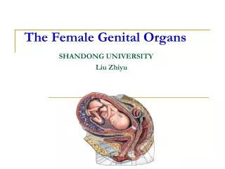
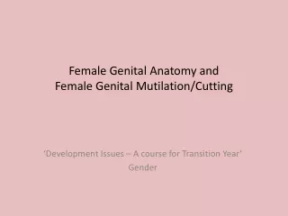
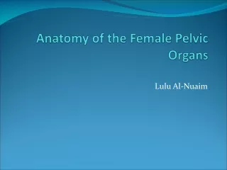
Characteristics of the female pelvis • Normal birth depends on: • The expulsive forces • The passages • The passenger The female pelvis is ”designed” to give room for the exit of the ”passenger”
Characteristics of the female pelvis, cont’d • Factors contributing to wider passages in the female : • Larger diameters of pelvic inlet • Less protruding promontory • Wider subpubic angle • Wider distance between the ischialtuberosities and the ischial spines • Widerand lower symphysis • Thesacrumcurvesless than in the male
Transverse diameter = 13 cm (Tverrvidden) Transverse diameter = 11 cm (intertubarvidden) Antero-posterior diameter =12,5 cm (diameter recta, likevidden) Antero-posterior diameter = 12 cm (conjugata vera, likevidden) Bispinous diameter = 10,5 cm (Interspinalvidden) The Diameters of the Birth Canal
Antero-posterior diameter = 12 cm (conjugata vera, likevidden) Antero-posterior diameter =12 cm (diameter recta, likevidden) The Bony Passages
Pelvic Fracture? Hamilton Bailey’s Physical Signs in Clinical Surgery Per Brodal 2008
Llewellyn-Jones 1999 Suboccipito-bregmatic diameter = 9,5 cm Anterior fontanelle (Bregma) Biparietal diameter = 9,5 The fetal skull Moulding during labor
Peritoneum Pelvic floor muscles (levator ani) Soft Parts of the Passages • Sheets of muscles and connective tissue • Several overlapping layers • Funnel-shaped • Stretched and pushed aside during childbirth • Subcutaneous and subperitoneal spaces with loose connective tissue, vessels, nerves and lymph nodes
Levator ani muscle Urogenital diaphragm with sphincter muscles Muscles of the pelvic floor Thieme, Atlas of Anatomy 2006
Formation of the Birth Canal and Moulding of the Fetus During Labour Amniotic fluid Soft tissues Skeleton