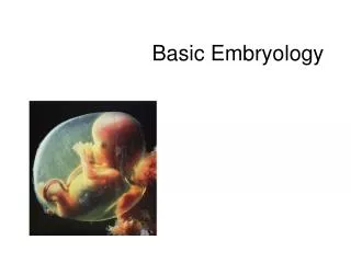
Basic Embryology
Basic Embryology. Embryology. Definition: the study of the origin and development of an organism Prenatal period: before birth 38 weeks from conception to birth (average) “fetal” age Gynecologic timing has been from LMP therefore refers to 40 weeks “gestational” age
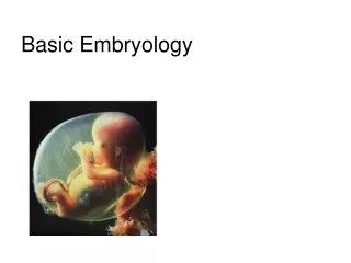

Basic Embryology
E N D
Presentation Transcript
Embryology • Definition: the study of the origin and development of an organism • Prenatal period: before birth • 38 weeks from conception to birth (average) “fetal” age • Gynecologic timing has been from LMP therefore refers to 40 weeks “gestational” age • Date of conception has been difficult to time • LMP is on average two weeks before ovulation
Traditional (artificial) division: • “Embryonic” period: first 8 weeks • All major organs formed • “Fetal” period: remaining 30 weeks • Organs grow larger and become more complex
Ovulation: egg released into the peritoneal cavity • Travels down fallopian tube in which fertilization occurs • At conception in fallopian tube, maternal and paternal genetic material join to form a new human life (zygote) • Cell division occurs with travel down the tube and into the uterus Conception (biology) or fertilisation, the fusion of gametes to produce a new organism of the same species (Wikipedia)
Week 1 post conception • Zygote divides repeatedly moving down tube toward uterus (cleavage) • The daughter cells are called blastomeres • Morula: the solid cluster of 12-16 blastomeres at about 72 hours • Day 4: late 60 cell morula enters uterus, taking up fluid becoming blastocyst
Blastocyst stage _____inner cell mass ______trophoblast • Two distinct types of cells • Inner cell mass: forms the embryo • Trophoblast: layer of cells surrounding the cavity which helps form the placenta • Floats for about 3 days • Implantation on about day 6 post conception • Trophoblast erodes uterine wall • Takes 1 week to complete • If inner cell mass of a single blastocyst divides: monozygotic (identical) twins
Week 2 • Inner cell mass divides into epiblast and hypoblast • 2 fluid filled sacs • Amniotic sac from epiblast • Yolk sac from hypoblast • Bilaminar embryonic disc: area of contact (gives rise to the whole body)
Blastocyst stage _____inner cell mass ______trophoblast • Two distinct types of cells • Inner cell mass: forms the embryo • Trophoblast: layer of cells surrounding the cavity which helps form the placenta • Floats for about 3 days • Implantation on about day 6 post conception • Trophoblast erodes uterine wall • Takes 1 week to complete • If inner cell mass of a single blastocyst divides: monozygotic (identical) twins
Week 3 • Bilaminar to trilaminar disc • Three primary “germ” layers: all body tissues develop from these • Ectoderm • Endoderm • Mesoderm
Formation of the 3 “germ” layers • Primitive streak (groove) on dorsal surface of epiblast • Grastrulation: invagination of epiblast cells • Days 14-15: they replace hypoblast becoming endoderm • Day 16: mesoderm (a new third layer) formed in between • Epiblast cells remaining on surface: ectoderm
The three “germ” tissues • “Germ” as in germinate, not germs • Early specialization of cells • Are precursors • Ectoderm and endoderm are epithelial tissue (form sheets of tissue) • Mesoderm is a mesenchyme tissue • Mesenchyme cells are star shaped and do not attach to one another, therefore migrate freely
Notochord • Days 16-18 • Primitive node epiblast cells invaginate and migrate anteriorly with some endoderm cells • Rod defining the body axis is formed • Future site of the vertebral column
Neurulation • Notochord signals overlying ectoderm • Formation begins of spinal cord and brain (neurulation) • Neural plate to neural groove to neural tube: pinched off into body
Closure of neural tube: begins at end of week 3; complete by end of week 4 (folic acid important for this step) • Extends cranially (eventually brain) and caudally (spinal cord) • Neural crest, lateral ectodermal cells, pulled along and form sensory nerve cells and other structures
Mesoderm begins to differentiate • Lateral to notochord, week 3 • Extends cranially and caudally (from head to tail or crown to rump) • Division of mesoderm into three regions • Somites: 40 pairs of body segments (repeating units, like building blocks) by end week 4 • Intermediate mesoderm: just lateral to somites • Lateral plate: splits to form coelom (“cavity”)
Divisions of the mesodermal lateral plate • Somatic mesoderm: apposed to the ectoderm • Splanchnic mesoderm: apposed to the endoderm • Coelom in between will become the serous cavities of the ventral body cavity: • Peritoneal • Pericardial • Pleural
Foldingbegins at week 4 (main difference between the 3 week embryo and the adult body is that the embryo is still a flat disc)
24 day embryro; protrudes into amniotic cavity
Day 23, beginning to fold Lateral folds will join ventrally
Cylindrical human body plan, day 28 (about ½ cm) Simplified cross section through abdomen of an adult (essentially the same as above)
29 day embryo(this is when the heart starts pumping, about 4 weeks or 1 month, ½ cm size)
month 3 month 5 3 month fetus (6 cm) late 5th month (about 19 cm)
By 8 weeks, about 2 months, all major organs are in place in at least a rudimentary form; this is why drugs early in pregnancy are so important to avoid – many cause birth defects; baby is a little over 1” long (below right)