Download
1 / 23
230 likes | 246 Vues
This study examines the diagnostic performance of Optical Coherence Tomography (OCT) in the non-invasive diagnosis of basal cell carcinoma (BCC). It analyzes the influence of lesion location, subtype, observer variability, and image quality on diagnostic accuracy.

E N D
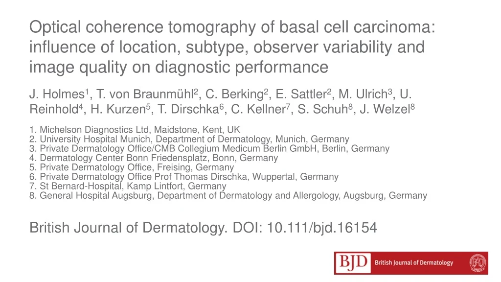
Optical coherence tomography of basal cell carcinoma: influence of location, subtype, observer variability and image quality on diagnostic performance J. Holmes1, T. von Braunmühl2, C. Berking2, E. Sattler2, M. Ulrich3, U. Reinhold4, H. Kurzen5, T. Dirschka6, C. Kellner7, S. Schuh8, J. Welzel8 1. Michelson Diagnostics Ltd, Maidstone, Kent, UK 2. University Hospital Munich, Department of Dermatology, Munich, Germany 3. Private Dermatology Office/CMB Collegium Medicum Berlin GmbH, Berlin, Germany 4. Dermatology Center Bonn Friedensplatz, Bonn, Germany 5. Private Dermatology Office, Freising, Germany 6. Private Dermatology Office Prof Thomas Dirschka, Wuppertal, Germany 7. St Bernard-Hospital, Kamp Lintfort, Germany 8. General Hospital Augsburg, Department of Dermatology and Allergology, Augsburg, Germany British Journal of Dermatology. DOI: 10.111/bjd.16154
Lead Author: Jon Holmes MA FInstP CEng CEO, Michelson Diagnostics Ltd
Disclosures Funding Sources This study was funded by Michelson Diagnostics Ltd (MDL), but they were not involved in the design or conduct of the study. The lead author of this paper is an employee of MDL who led the analysis of the data under the supervision of the final author. Conflicts of Interest J.H. is an employee and shareholder of Michelson Diagnostics Ltd which manufacturers OCT equipment used in the study. M.U. has been paid for lectures for Michelson Diagnostics Ltd; is a stakeholder in CMB Collegium Medicum Berlin GmbH; has been involved in clinical trials for LEO Pharma and has performed paid lectures for Almirall, Galderma, LEO Pharma, and Mavig GmbH. T.B. has received speaker’s honoraria from Agfa HealthCare GmbH, LEO Pharma, and Roche Pharma; and has been involved in clinical trials sponsored by Agfa HealthCare GmbH and Mavig GmbH. H.K. is a paid advisor to Michelson Diagnostics Ltd; has received speaker’s honoraria from Michelson Diagnostics Ltd, AlmirallHermal, AbbVie, Galderma, and LEO Pharma; and has been involved in clinical trials for Eli Lilly Pharma, Novartis and AbbVie. T.D. has received advisory board honoraria from AlmirallHermal, Biofrontera, Galderma, Meda Pharma, Scibase, and Allergan; and has received speaker’s honoraria from AlmirallHermal, Biofrontera, Galderma, Meda Pharma and Janssen- Cilag. C.B. has received speaker’s and advisory board member’s honoraria from Almirall, Biofrontera, Galderma, and LEO Pharma; and has been involved in clinical trials sponsored by Biofrontera and LEO Pharma.
Introduction What’s already known? • Optical Coherence Tomography (OCT) is an emerging imaging modality that has been shown to have utility in the non-invasive diagnosis of basal cell carcinoma and is more sensitive and more specific than clinical or dermoscopic examination alone.
Objective • What does this study add? • Lesion anatomical location does not affect diagnostic performance with OCT. • Poor OCT image quality is associated with superficial scales and crusting, reducing diagnostic performance; but in these cases diagnosis with OCT is better than by clinical or dermoscopy examination alone.
Objective (2) • Observers’ diagnostic confidence increases when using OCT, and their performance reflected this. • Diagnostic performance is consistent between trained observers. • BCC subtype can be diagnosed from OCT images with moderate accuracy.
Methods: Overview • This study quantified diagnostic performance of OCT imaging for the diagnosis of BCC vs. clinical assessment and dermoscopic examination, verified by histology • The study was: • Investigator led • At 6 centres, based in Germany • N=234 lesions suspicious for BCC • Prospective, Observational, Phase IV • Conducted in 2014-15
Protocol VivoSight OCT scanners were used to scan all lesions
Information captured (1) • For each lesion, and for each stage of observation (clinical, dermoscopy, OCT), the observer recorded: • Their diagnosis of the lesion (including BCC subtype) • Anatomical location of the lesion • Confidence level in the diagnosis
Information captured (2) • Additionally, for the OCT observation: • Assessment of OCT image quality (poor / mediocre / good /excellent) • Presence or absence of 15 image ’biomarkers’ which may be diagnostically useful
Results: Diagnostic Performance These results were first published in British J. Dermatology in 2015: Ulrich, M., T. Braunmuehl, H. Kurzen, T. Dirschka, C. Kellner, E. Sattler, C. Berking, J. Welzel, and U. Reinhold. "The sensitivity and specificity of optical coherence tomography for the assisted diagnosis of nonpigmented basal cell carcinoma: an observational study." British Journal of Dermatology 173, no. 2 (2015): 428-435.
Results (1) • In this additional 2017 research presented here, the 2014/15 dataset was analyzed to extract further useful results: • Anatomical location (trunk / head /limb ) did not effect the diagnostic performance of any method • NPV improved from 83% with mediocre OCT image quality (N=58) to 97% with good or excellent OCT image quality (N=161)
Results (2) • NPV improved from 90% with low/medium observer confidence level (N=105) to 98% with high/very high confidence level (N=129) • NPV was within range 88-100% for all 6 observers
BCC Subtype diagnosis and image biomarker analysis Key: (+) weak positive correlation (++) strong positive correlation (-) weak negative correlation (--) string negative correlation 62% - 72% of BCC subtypes were correctly identified with OCT
Discussion (1) • This new 2017 analysis of the 2014/2015 dataset has revealed additional information of value to the clinician interested in using OCT for non-invasive BCC diagnosis • Reduced OCT image quality was associated with superficial scales/ crusting (although performance with OCT was still better than by clinical or dermoscopic examination)
Discussion (2) • Observer performance correlated with their own confidence in their diagnosis. Therefore, it may be useful to decide on further actions (eg. taking a biopsy) based on observer confidence. Furthermore, observer confidence may be a useful marker for capturing and documenting observer expertise with OCT
Discussion (3) • Observer performance was consistent across all 6 sites • There were clearly differences in the markers observed for the 3 different BCC subtypes (and also for the non-BCC lesions) • The findings demonstrate that OCT usefully visualizes the histopathology of architectural features of BCCs – in contrast to clinical and dermoscopic examination which cannot do this
Discussion (3) - Limitations • Dataset was limited to 234 lesions, which is small for analysis of subtype and did not include all possible non-BCC lesion types • All 6 observers were experienced in non-invasive imaging and use of OCT; therefore not representative of new OCT users • Some results were subjective (presence of some image markers; confidence level; image quality) • Histology, used as the gold standard in this study, may not be 100% accurate (sampling error of biopsy)
Conclusions (1)What does this study add? • OCT improves the differential diagnosis of BCC versus other lesion types in clinically suspicious lesions compared to clinical and dermoscopic diagnosis alone as reported previously; • There are a number of useful ‘image biomarkers’ that aid the OCT user in diagnosing BCCs versus other lesion types, but further research is needed to find additional new independent markers;
Conclusions (2) • Poor OCT image quality is associated with superficial scales and crusting, and this impacts diagnostic performance using OCT; however diagnosis aided by OCT in these cases is still better than by clinical or dermoscopy examination alone;
Conclusions (3) • Observers’ own confidence in their diagnosis of BCC increased when using OCT versus clinical and dermoscopy alone, and their actual diagnostic performance reflected this (i.e., they were more likely to be right when they had high confidence); • Observer diagnostic performance was consistently better with OCT than with clinical examination or dermoscopy alone across all test sites.
Research Team J Holmes T. von Braunmühl C. Berking E. Sattler M. Ulrich U. Reinhold H. Kurzen T. Dirschka C. Kellner S. Schuh J. Welzel
Call for correspondence • Why not join the debate on this article through our correspondence section? • Rapid responses should not exceed 350 words, four references and one figure • Further details can be found here