Download
1 / 27
550 likes | 2.19k Vues
Autonomic Nervous System (ANS). Introduction to ANS: It is the part of the peripheral nervous system (Cranial and spinal nerves ). It composed of nerves, ganglia , and plexuses coordinates the function of internal organs of the body.

E N D
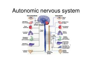
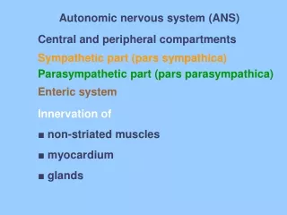
Autonomic Nervous System (ANS) Introduction to ANS: It is the part of the peripheral nervous system (Cranial and spinal nerves). It composed of nerves, ganglia, and plexuses coordinates the function of internal organs of the body. It regulates respiration, circulation, digestion, metabolism, reproduction, and many other function of the body. These functions are also maintain by this system during sleep and in general anesthesia, hence the term autonomic nervous system designates.
Autonomic Nervous System (ANS)(cont.) Introduction to ANS: • The life sustaining activities such as visceral (internal soft organ) function and homeostasis (internal physiology) which is out of individuals controlled is maintain by the autonomic nervous system. • The visceral function include also the secretion of endocrine and exocrine function and the function of blood vessels also. • The ganglia include paravertebral chain ganglia and prevertebral ganglia, and intramural ganglia.
Difference between Somatic Nervous System and Autonomic Nervous System (1) Somatic Nervous System (SNS), is under voluntary control, and regulates the contraction of skeletal muscle. In contrast, the autonomic nervous system (ANS) or visceral motor system, is generally not under voluntary control and controls smooth muscle, cardiac muscle, glandular tissue and visceral reflexes.
Difference between Somatic Motor System and Autonomic Nervous System(cont.) (2) In the SNS, efferent fibers (motor nerve) begins from the cortex of brain, and travel either to the cranial nerve nuclei located in the brain or to the alpha-motor neuron of the dorsal horn of the spinal cord. From here, motor nerves exit the CNS and innervates into the skeletal muscle. Therefore, in somatic nervous system the nerve cell bodies of the motor nerves located in the CNS.
Difference between Somatic Motor System and Autonomic Nervous System(cont) (2) In the ANS, efferent fibers (motor nerve) begins from the hypothalamus of brain and travel to the nuclei of the cranial nerves belong the ANS or intermediate horn of the spinal cord. From here the nerves (preganglionic fibers) travel again and synapse in the 2nd ganglion (paravertebral, prevertebral, or intramural ganglia) located outside the CNS.
Difference between Somatic Motor System and Autonomic Nervous System(cont) 3. The fibers of SNS innervates into the skeletal muscle as neuromuscular spindle. Whereas the ANS innervates into the smooth muscle as varicosities and vesicles.
Division of Autonomic Nervous System Based on anatomy, physiology, and pharmacology, the ANS can be divided into: (1) Sympathetic division associated with autonomic nerves, paravertebral chain ganglia and prevertebral ganglia. (2) Parasympathetic division, associated with autonomic nerves and intramural ganglia.
Characteristics of the Fibers of Autonomic Nervous System (ANS) • The fibers are: Preganglionic & postganglionic fibers. • The autonomic fibers from the intermediated horn of the spinal cord to the paravertebral or prevertebralganglionic synapse is called preganglionic fibers are myelinated, therefore, it is white. • The postganglionic fibers are the fibers after the pre- or paravertebral ganglia which are little myelinated, therefore, it is gray.
Structural Organization of Sympathetic division • In sympathetic division the multipolar nerve cell body is located in the intermediate horn of the thoracic and lumbar spinal cord (between 1st thoracic and 3rd lumbar segment). • The preganglionic nerve fiber (myelinated axon) is short and travel from the multipolar nerve cell body and synapse within the paravertebral chain ganglia/ prevertebral ganglia. • The postganglionic fiber (non-myelinated) is long and terminates on the cells of effector organ.
Nerve cell bodies located in the Paravertebral and Prevertebral ganglion Intermediate horn Paravertebral Ganglion Prevertebral Ganglion
Autonomic Nerve Fibers Preganglionic nerve fiber Preganglionic nerve fiber Postganglionic nerve fiber Synapse here in The paravertebral Ganglion Postganglionic nerve fiber Synapse here in the Prevertebral ganglion
Review of Ganglia and Nerves of Sympathetic Division Autonomic fibers from Intermediate horn of spinal cord Intermediate horn White rami Myelinated Preganglionic fiber Gray rami Ventral root of thoracic and 1st few lumbar spinal nerve PVG Non-myelinated Postganglionic fiber Prevertebral Ganglion (PrVG) White rami Synapse in the paravertebral Ganglia/prevertebral ganglia (This is the preganglionic fiber) The postganglionic fiber from here innervates into the autonomic organ
Sympathetic Trunk Sympathetic trunk consist of paravertebral ganglia on either side of the vertebral column and are interconn -ected longitudinally and transversely. It can be divided into: • Cephalic and Cervical Part • Thoracic Part • Abdominal part and • Sacral and Coccygeal part.
Cephalic and Cervical part of Sympathetic Trunk • It is the cranial continuation of the thoracic part, without directly contacting the vertebral column. • The cervical part starts at the cervico-thoracic ganglion, which is connected to the middle cervical ganglion. From here sympathetic trunk passes cranially in combination with the vagus nerve forming “vagosympathetic trunk”. At the atlas, the sympathetic part separates from the vagus and terminate into cranial cervical ganglion.
Ganglia and autonomic innervation in the Cervical Trunk • The cranial cervical ganglion is located near the atlas bone and serves for the autonomic innervation of the head and face. • Middle cervical ganglion is located close to the subclavian artery and serves for the autonomic • innervation of the heart.
Ganglia and autonomic innervation in the Cervico-thoracic region • Cervicothoracic ganglion: The cervico -thoracic ganglion is located medial to the first rib. It is the results of the fusion of caudal cervical and one or two thoracic ganglion. It send following branches: • Vertebral nerve • Cervical cardiac nerve,& • Caudal continuation of thoracic sympathetic part.
Thoracic part of the sympathetic trunk (See Fig. Slide 14) • The number of paravertebral ganglia is correspond to the number of thoracic vertebra. • The organs of the thoracic cavity (heart, lungs, trachea received autonomic innervations from Postganglionic fibers originating from cervicothoracic ganglion or from postganglionic thoracic nerves. • More caudally 7-8 preganglionic thoracic fibers unite to form greater splanchnic nerve pass through the crus of diaphragm, entered into the abdominal cavity and synapse in the celiac ganglia. The postganglionic fiber from here supply to the liver, stomach, spleen, and pancreas. • Last 2-3 thoracic preganglionic fibers unite to form lesser splanchnic nerve, enter into the abdominal cavity and synapse in cranial mesenteric ganglion. The postganglionic fibers from here supply to the jejunum, ileum, cecum, colon, and kidney.
Abdominal part of the sympathetic trunk (See Fig. Slide 14) • The lumbar autonomic nerves leaves the lumbar segments of spinal cord, pass through the paravertebral ganglia and forms lumbar splanchnic nerves, and synapse with the celiac ganglion, cranial mesenteric ganglion, and caudal mesenteric ganglion. The postganglionic fibers innervates into spleen, kidney, pancreas, cecum, colon, rectum, and genital organs.
Sacral part of the Sympathetic Trunk (See Fig. Slide 14) First 5 preganglionic nerve fibers leaves the pelvic part of the spinal cord, pass through the paravertebral ganglion, unite to form sacral (pelvic) spalnchnic nerves which synapse in the sacral ganglion (pelvic ganglion). The postganglionic fibers innervates into the rectum and genital organs.
Structural Organization of Parasympathetic division • The multipolar nerve cell body is located in the nuclei of the III, VII, IX, and X cranial nerves and intermediate horn of first 3-5 sacral spinal cord of different animals. • The preganglionic nerve fiber (myelinated axon) is long and travel from the multipolar nerve cell body and synapse within the intramural ganglia; ciliary, pterigopalatine, mandibular, otic and distal ganglia. • The postganglionic fiber (non-myelinated) is short and terminates on the cells of effector organ.
Parasympathetic Innervation in the Head Region Occulo. nerve III. Oculomotor nerve Preganglionic fiber synapse In the ciliary ganglion Postganglionic fiber innervates into ciliary muscle which regulates lens curvature and the pupillary diameter.
Parasympathetic Innervation in the Head Region Facial n. VII. Facial nerve Preganglionic fiber synapse in the Pterygopalatine and mandibular ganglion Postganglionic fiber innervates into sublingual, submandibular, lacrimal, nasal, and palatine gland.
Parasympathetic Innervation in the Head Region G.Pharyn. nerve IX. Gloss. Phar.nerve Preganglionic fiber synapse in the Otic ganglion Postganglionic fiber innervates into buccal and parotid gland.
Parasympathetic Innervation of the Vagus Nerve Vagus Nerve Vagus nerve Preganglionic fibers innervates into the organ of neck, thoracic abdominal and pelvic region and synapse with the intramural ganglion located in the organs. Postganglionic fiber is short. Vagus n in the Abdomen Cervical Vagus n. Vagus n in the Pelvic region Vagus n. in the Thoracic region
Pelvic Parasympathetic Innervation Preganglionic fibers from intermediate horn of the spinal cord forms pelvic autonomic nerves and innervates into the pelvic organs and synapse with the intramural ganglion of the organs. Postganglionic fibers are short. Pelvic Parasym- pathetic nerves
Function of Autonomic Nervous System in General Both the system have opposing effects on most function: • Sympathetic (Thoraco-lumbar nerves) division generally causes excitation and results in catabolism. This division is acted during period of stress and exertion. Sympathetic nerve ending secretes neurotransmittor (chemicals) “norepinephrine”. Norepinephrine act on α, and β adrenergic receptor of various organ, that is why sympathetic nerves is called Adrenergic nerves. • The parasympathetic division is responsible for rest, digestion, and anabolism (i.e., building phase of metabolism). Parasympathetic nerve fibers secrete neurotransmittor acetylcholine, that’s why these nerves known as Cholinergic nerves.
Function of Autonomic Nervous System(Specific) Organ Sympathetic Parasympathetic Nervous System Nervous System 1.Iris Dilates pupil Constrict pupil 2. Ciliary m. Relax Constrict 3. Glands Inhibit secretion Stimulates secret. 4. Heart m. Increased HR Decreased HR 5. U. Bladder Inhibit urine release Cause urination 6. Lung Dilates bronchioles Constrict Br. 7. Digestive tract Decreased peristalsis Increased