Download
1 / 34
340 likes | 376 Vues
226 PHT Lab # 1. Basic Microbiology. Microbiology is the science that deals with the study of too small organism (micro - organisms) that are invisible to the naked eye. Laboratory Rules and Safety. Instructions and Rules. ☠ No eating or drinking. ☠ Lab coat & marker.

E N D
226 PHT Lab # 1
Basic Microbiology Microbiology is the science that deals with the study of too small organism (micro-organisms) that are invisible to the naked eye
Instructions and Rules ☠ No eating or drinking. ☠ Lab coat & marker. ☠ Aseptic technique. ☠ Benches must be disinfected. ☠ ☠ Avoid contamination. (Discarded cultures & infectious materials and broken or spilled living cultures).
☠ Microscope. ☠ At the end of each lab check: • Gas tap is turned off. • Water tap is closed properly. • Microscope lamp is turned off. ☠ Finally wash your hands thoroughly.
Introduction to Microbiological Equipments and Materials • Medium:- ❊An artificial preparation contains the essential elements and nutrients needed by the m.o to grow (media)
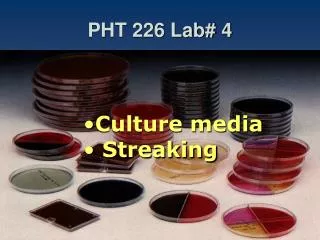
Culture media ❊It may be: • Liquid (broth) • Solid (containing agar) • Semisolid (containing low conc. of agar)
Introduction to Microbiological Equipments and Materials • Inoculation: Culturing of sterile media with m.o [Inoculation loop]. • Incubation: Placing the culture into the incubator at optimum temperature for growth. • Sterile: Free from any living microorganism • Contamination: Introduction of undesirable m.o.
Sterilization: Killing or removal of all living micro-organisms (from a particular location or material). Sterile article:completely free of all living micro-organisms Disinfection: Destruction of vegetative conspiring micro-organisms. .
Disinfectants: • Chemicals which cause disinfection • Bacterial spores, mycobacteria, some viruses → considerable resistance • Antiseptics: • Disinfectants which can be safely applied to skin & mucous membranes.
Contamination: • Introduction of undesirable m.o. • Asepsis: • Processes designed to prevent m.o. from reaching a protected environment. • Aseptic technique: Practices used by microbiologists to exclude all organisms from contaminating media or contacting living tissues.
Discard cultures and other infectious materials: • Petri dishes→ Plastic bag → Autoclave. • Test tube cultures → wire basket → Autoclave. • Used pipettes → Plastic bag → Autoclave. • Used slides, covers, and pipettes → Jar containing a disinfectant. • Broken glass → swept in a dustpan → container for broken glass. NEVER place contaminated material in waste basket.
Broken or spilled living cultures: • Clothing → Autoclave plastic bag → Autoclave. • Flood the area with a disinfectant ( or paper towels are placed over the spills). • After 20- 30min→ wipe up & discard the waste in autoclavable dustpan→ Autoclave.
Identification of Bacteria • Microscopical Examination: • Characters of cells. • Macroscopical Examination: • Characters of colonies.
Identification of Bacteria • Biochemical Tests. • Additional Tests: • such as seriological tests
Colony vs. Cell Cells Colonies
Colony vs. Cell Cells Colonies
The Microscope • The microscope is the most important tool used for examination and identification of microorganisms.
The Microscope Uses of microscope: • Identification of microbial groups. (Bacterial, Fungi, Protozoa) • Morphological studies of m.o (size, shape, arrangement…..) 3. Physiological studies (motility ….)
The Microscope Types of microscopes: • Optical microscope: • Use light beams and lenses • The most common one used in the lab 2. Electronic microscope: • Use electron beams and magnetic fields • Used for examination of viruses and sections of bacteria
Optical microscope vs. Electronic microscope Optical microscope Electronic microscope
Optical microscope vs. Electronic microscope Electronic microscope Optical microscope
Optical microscope vs. Electronic microscope Optical microscope Electronic microscope
The Microscope • Optical microscope: • There are two types a- Simple microscope: single system of lenses b-Compound microscope: has two lens system, the ocular lens and the objective lens • The two lenses system give greater magnification
Components of the compound microscope Ocular lens Objective lens Stage Iris diaphram Lamp box Slide movement knob Fine adjustment Coarse adjustment
Theoretical principles of microscopy Magnification: • It means enlargement of the linear diameter of an object. • It is the function of two lens system [the ocular and the objective lens]
Theoretical principles of microscopy • Total magnification = mag. power of ocular lens X mag. power of objective lens used N.B (oil immersion lens is used) Objective lens mag. Power • Scanning lens 4× • Low power objective lens 10× • High power objective lens 40× • Oil immersion lens 100×
Theoretical principles of microscopy • The magnification power of ocular lens (eye piece) is 10× • The total magnification will be 40, 100, 400, 1000 times.
Theoretical principles of microscopy • Working distance: It is the distance between the objective lens and the slide. • General rule, as the magnification of the lens increase the working distance decrease.
Theoretical principles of microscopy • Resolution: It is the ability of a lens to reveal two closely adjacent points as separate and distance. • The resolving power of the oil immersion lens depends on the addition of special oil (Ceder wood oil) between the specimen slide and the objective lens.
Resolution: Higher Resolution; note the “Sharpness” of the Image. Which image is least resolved? Lower Resolution; it is less sharp.
Oil Immersion Increases Resolution Air has a different Index of Refraction from water (so light bends). Air has a different Index of Refraction from glass (so light bends). The Mineral Oil has the same Index of Refraction as glass (so light does not bend).
Objective lens Lost light Saved light oil specimen Light source