Plantar Fasciitis
420 likes | 1.64k Vues
Plantar Fasciitis. Michael LaBella. Objectives. Learn the anatomy of the foot. Identify key terms associated with plantar fasciitis. Determine the causes of plantar fasciitis and understand why it occurs. Recognize the injury when it occurs and be able to evaluate plantar fasciitis.

Plantar Fasciitis
E N D
Presentation Transcript
Plantar Fasciitis Michael LaBella
Objectives • Learn the anatomy of the foot. • Identify key terms associated with plantar fasciitis. • Determine the causes of plantar fasciitis and understand why it occurs. • Recognize the injury when it occurs and be able to evaluate plantar fasciitis. • Learn about current treatments as well as the rehabilitation for plantar fasciitis. • Be able to understand the methods available for the prevention of plantar fasciitis.
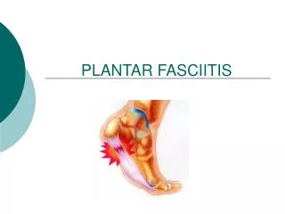
Anatomy of the foot • The plantar fascia is a ligament structure that supports the longitudinal arch of the foot. It is a tough, fibrous band of connective tissue that runs from the heel bone to the ball of the foot. The plantar fascia is made up of predominantly longitudinally collagen fibers. In the ligament, there are three distinct structural components, the medial, central and the lateral component. The central component is the largest and most prominent.
Anatomy of the foot • The foot consists of 26 bones that form two arches, one longitudinal and one lateral..
Anatomy of the foot • As stated before, there are 26 bones, 33 joints, 106 ligaments, 19 foot muscles and 11 muscles in the lower leg. The foot enables us, through subtle movements, to walk and run up to 10,000 to 17,000 steps a day. During our lives our feet carry us between 65,000 and 115,000 miles.
Anatomy of the foot (con’t) • The arches of the foot function as shock absorbers, supporting the body and enabling stable ambulation. Clinicians typically divide the foot into three zones: the forefoot, midfoot, and hindfoot. The plantar fascia is a dense, fibrous membrane that spans the entire length of the foot, originating at the tubercle of the calcaneus and attaching at the proximal phalanges. The fascia protects the underside of the foot and helps support the arches. It consists of a thick central portion and thinner lateral and medial bands that provide flexibility and help maintain the longitudinal arch
Key terms • Plantar Fasciitis: an inflammation caused by excessive stretching of the plantar fascia.
Key terms (con’t) • Plantar Fascia: a broad structure that spans between the medial calcaneal tubercle and the proximal phalanges of the toes. It is the thick connective tissue which supports the arch of the foot.
Key terms (con’t) • Dorsiflexion: the movement which decreases the angle between the foot and the leg. • Plantarflexion: the movement which increases the angle between the foot and the leg. • Pronation: a rotation movement, the foot pronation will cause the sole of the foot to face more laterally than when standing in the anatomical position.
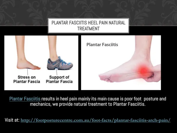
Why does plantar fasciitis occur, and to whom? Plantar fasciitis is common in middle-aged men and women, but can be diagnosed in all age groups. It occurs in people who tend to stand for long periods of time, or with people who have gained weight rapidly. With athletes, runners are most susceptible due to the repetitive act of foot flexion while running. All these factors, including the wearing of shoes with little or no arch support, and inactivity are also associated with the condition.
Why this injury occurs • Plantar fasciitis occurs due to irritation to the ligamentous connective tissue. The inflammation in the tissue is the result of some type of injury to the plantar fascia. Typically, plantar fasciitis results from repeated trauma to the tissue where it attaches to the calcaneus. This may result in the tearing of the plantar fascia at or near the point of attachment of the tissue to the calcaneus. The result of the damage and inflammation is a dull, aching pain under the foot.
Why this injury occurs (con’t) • People who develop plantar fasciitis have several risk factors, they include: • Flat feet • High arched, rigid feet • Increasing age and family tendency • Running on toes, hills or very soft surfaces (sand) • Poor arch support in shoes • Rapid change in activity level
Evaluation and symptoms of Plantar Fascitiis • Patients usually describe pain in the heel on taking the first several steps in the morning, with the symptoms lessening as walking continues. • Many patients believe the condition to be the result of recent increase in daily activity. It is not unusual for a patient to endure the symptoms many years before seeking medical treatment.
Evaluation (con’t) • The person affected by this injury often gives a history of a gradual onset of pain which is worst on first weight bearing in the morning. After a few steps, the heel pain will decrease during the day but will worsen with increased activity (such as jogging) or after a period of sitting. Worse pain in the morning is typical of plantar fasciitis. Nighttime pain should raise the suspicion of other causes of heel pain such as tumors, infections, and neuropathic pain. • The patient may describe an aggravating factor with the discomfort gradually increasing over subsequent weeks. An accurate history of footwear should be obtained: often patients wear shoes with poor cushioning or inadequate arch support, or they walk barefoot on hard floors.
Examination of Plantar Fascitiis • Physical examination in a patient with plantar fasciitis shows tenderness on the heel; firm finger pressure is often necessary to localize the point of maximum tenderness. Slight swelling in the area is common. Tightness of the Achilles tendon is found in 78% of patients.
Types of evaluation conducted Lateral radiograph- of the heel is usually taken to rule out a stress fracture or rare bony causes of inferior heel pain. • Isotope scanning– Isotope scanning is of questionable value, and later the diagnosis can usually be made clinically; scanning should be used only when stress fractures are suspected and plain radiographs are normal. Magnetic resonance imaging and ultrasonography– Magnetic resonance imaging is rarely indicated but may show thickening and inflammation of the fascia. Ultrasound examination too may show increased thickness of the plantar fascia and appearances of inflammatory changes.
Types of evaluation conducted (cont’d) • Blood tests– A full blood count and erythrocyte sedimentation rate are recommended. • Electrophysiological studies– The tarsal tunnel syndrome may sometimes be confirmed, but the studies are difficult to perform and interpret.
Treatment of Plantar Fascitiis • Initial treatment within the first 48 hours for plantar fasciitis can be as simple as limiting one to rest, or can be as aggressive as seeking medical attention. (MRI’s, inflammatory or pain relievers) Plantar fasciitis rehabilitation time can be up to six to eighteen months. Rest is the number one prescribed treatment to plantar fascitiis. However, it is equally important to correct the problems that place individuals at risk, such as increased amount of weight-bearing activity, increased intensity of activity and increased walking activities.
Treatment of Plantar Fascitiis • Stretching and StrengtheningStretching and strengthening programs play an important role in the treatment of plantar fasciitis. Increasing flexibility of the calf muscles is particularly important. Frequently used stretching techniques include wall stretches and curb or stair stretches.
Treatment of Plantar Fascitiis • The Slant-board Stretch & the Stair stretch
Treatment of Plantar Fascitiis (con’t) • Dynamic stretches such as rolling the foot arch over a can or a tennis balls are also useful. Cross-friction massage above the plantar fascia and towel stretchingmay be done before getting out of bed.
Treatment of Plantar Fascitiis (con’t) • Treatment of plantar fasciitis consists of 3 stages: • Stage 1 involves prescribing the patient a heel cushion to decrease shock absorption of the plantar fascia, as well as a short course of medication to decrease inflammation in the heel. • Stage 2 involves cortisone injections into the heel region, if Stage 1 has failed to bring significant relief. Other modalities in Stage 2 include: orthotics, taping, physical therapy, and night splinting. • Stage 3 is for those who have had plantar fasciitis for one year or longer, whose symptoms are severe and preventing them from their job or recreation. It involves a surgical release of part of the insertion of the plantar fascia. However, this surgery is rare, as most people do have significant relief from non-surgical treatment.
Treatment • Some other treatments available are supports such as night splints which stretch the plantar fascia while you sleep. By stretching the plantar fascia, it will be forced to lengthen and avoid the early morning tightness and pain brought on by lack of use overnight while at rest.
Rehabilitation • Rest from activities that cause pain. Stay off your feet as much as you can. Use crutches if necessary. Maintain fitness by swimming or cycling. Take the opportunity to work on upper body strength. • If you cannot stay off your feet then the next best thing is to tape the foot. The taping gives excellent support while allowing the foot to heal. If taping is effective then it is likely that orthotics will also be effective in correcting foot biomechanics and helping to prevent the injury returning once normal training has resumed. • Apply cold therapy. Ice massage for 10 minutes to the site of pain - every hour for the first day progressing to three times a day at least. • NSAID (Non steroidal anti-inflammatory medication) like ibuprofen may help in the early stages.
Rehabilitation (con’t) • PHASE 1- Acute Phase: • Goal decrease acute pain and inflammation: • absolute or relative rest- Decrease sports activity to avoid rebound pain • ICE: 2 applications of 20 minutes per day • Non steroidal aids
Rehabilitation (con’t) • PHASE 2- Rehabilitation Phase: • Further decrease pain and inflammation: • ultrasound • phonophoresis • neuroprobe • contrast baths • Maintain/increase flexibility of injured (and surrounding) tissue: • gentle stretching exercises: calf, hamstring, posterior muscle groups
Rehabilitation (con’t) • PHASE 3- Functional Phase: • Functionally strengthen intrinsic muscles of the foot • closed chain therapeutic exercise • Doming of Arch (towel toe curl) • Protect injured area during functional activity • taping • stability running or other appropriate athletic shoes • orthoses as needed
Returning to play • When you have gone a week with no pain then you can begin to run again. • This should be a gradual process. If you feel pain at any time then go back a step. • Running time should be gradually increased. • Apply tape to the foot to support it for the first few runs, especially if you do not have orthotics. • Ensure you have the correct shoes for your running style or sport. • After every training session apply ice to the shin for about ten minutes. • Ensure you stretch properly before each training session and after. Hold stretches for about 30 seconds and repeat 5 times.
Returning to play (con’t)Below is an example of a gradual return to running. Begin each training session with a 5 minute walk followed by a stretch Day 1: walk 4 minutes jog 2 minutes repeat four times • Day 2: rest • Day 3: walk 4 minutes jog 3 minutes repeat three times • Day 4: rest • Day 5: walk 3 minutes jog 4 minutes repeat 4 times • Day 6: rest • Day 7: walk 2 minutes jog 6 minutes repeat 4 times
Prevention • The following steps will help prevent plantar fasciitis or help keep the condition from getting worse if you already have it • Take care of your feet. Wear shoes with good arch support and heel cushioning. • Do exercises to stretch the Achilles tendon at the back of the heel. This is especially important before sports, but it is helpful for non-athletes as well. • Stay at a reasonable weight for your height. • Establish good exercise habits. Increase your exercise levels gradually, and wear supportive shoes. • If you run, alternate running with other sports that will not cause heel pain. • Put on supportive shoes as soon as you get out of bed. Going barefoot or wearing slippers puts stress on your feet.
References • American Academy of Podiatric Sports Medicine. Plantar Fasciitis: Treatment Pearls2003 Douglas Richie, Jr. D.P.M. http://www.aapsm.org/plantar_fasciitis.html • The Physician and Sports Medicine Plantar Fasciitis Vol 32 number 11 2004 http://www.physsportsmed.com/issues/2004/1104/glazer.htm • Journal of Athletic Training Plantar Fasciitis: Heel Pain in Athletes. Jeffery A. Middleton, MS,. Eric L. Kolodin, DPM Volume 27 Number 1 . 1992 http://www.pubmedcentral.nih.gov.ezproxy.hsclib.sunysb.edu/picrender.fcgi?artid=1317134&blobtype=pdf • Southern California Orthopedic Institute Plantar Fasciitis Southern California Orthopedic Institute Jonathan S. Jaivin, MD 2004. http://www.scoi.com/plantar.htm