
Cardiovascular System: Anatomy, Physiology, and Disease
This chapter provides an introduction to the cardiovascular system, including its components, functions, and common pathologies such as heart failure and myocardial infarction. Learn about the structure and function of the heart, the route of red blood cells, and the measurements used to diagnose a heart attack.

Cardiovascular System: Anatomy, Physiology, and Disease
E N D
Presentation Transcript
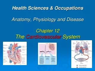
Health Sciences & OccupationsAnatomy, Physiology and Disease Chapter 12The Cardiovascular System
Introduction Cardio-vascular system • Transportsnutrients & oxygen to cells in body while carbon dioxide and waste products of cells’ metabolism are removed. • Pump that circulates the transport medium (blood) is the heart.
Components include heart, blood, and network of blood vessels. Arteries carry blood away from heart, branch into smaller vessels called arterioles, which become capillaries, where nutrients are exchanged; capillaries become venules, that enlarge and become veins. System Overview
System Overview con’t • Veins differ from arteries because they carry blood toward heart, have valves, and have thinner walls.
The Heart • Size of your fist: located slightly left of center of chest. • Base isproximal to your head while apex is distal. • One single organ but with two pumps working together. • Right side collects blood from body and sends it to lungs; left side collects blood from lungs and sends it to rest of body.
Four Chambers in Heart Septum: seperates heart in right & left half • Interatrial Septum: seperates right Atrium from left Atrium. • Interventricular Septum: seperates right & left ventricles.
Chambers of the Heart con’t • Right Atrium: collecting chamber where blood is returned to heart after trip around body. • Superior & Inferior Vena Cavae: large veins return blood to right atrium. • Tricuspid Valve (atrioventricular): directs blood from rt. Atrium to rt. Ventricle.
Chambers of the Heart con’t • Heart contraction occurs: when rt. Ventricle is full of blood. • Tricuspid valve prevents backflow of blood into rt. Atrium. • Blood flows through pulmonary semilunar valve to “pulmonary arteries.”
Chambers of the Heart con’t • Rt. & Lt. “pulmonary arteries” goes to lungs where vessels get smaller and smaller, ending in capillaries around each air sac (alveolus) • Blood returns to Lt. Atrium via “pulmonary veins.”
Chambers of the Heart con’t • Mitral Valve (Bicuspid Valve): allows blood flow from Lt. Atrium to Lt. Ventricle. • Left ventricular pressure increases as it fills • Heart contracts forcing mitral valve (Bicuspid Valve) closed. • Blood is ejected through aortic semilunar valve to ascending aorta, and then out torest of body.
The Route of an RBC • Superior/Inferior vena cava • Right Atrium • Tricuspid valve • Right Ventricle • Pulmonary Valve • Pulmonary Artery • Lungs • Pulmonary Veins • Left Atrium • Bicuspid Valve • Left Ventricle • Aortic Valve • Aorta
Chambers of the Heart con’t • Systole: contraction phase when blood is ejected from the ventricles. • Contraction: begins at apex and travels upward • Diastole: resting period when chambers refill with blood.
Chambers of the Heart con’t • Atrial walls: thinner than ventricular walls Ventricular walls: • Lt. ventricle pumps blood to body thus thick walls • Rt. Ventricle pumps blood to lungs thus thinner walls.
Coronary Arteries • Right coronary artery: provides blood for right ventricle, posterior portion of interventricular septum, and inferior parts of heart. • Left coronary artery: provides blood to left lateral and anterior walls of left ventricle, and portions of right ventricle and interventricular septum.
Pathology Problems (CHF) • Rt. SideHeart Failure:(corpulmonale) Etiology: muscles chronically work harder than normal resulting in large muscle & inefficient pumping; pulmonary embolus, COPD. S/S: SOB, wheezing, engorged liver & spleen, ankle, feet & hand edema, & JVD (distended neck veins) Dx: Chest X-ray Rx: O2, diuretics, digitalis, nitrates, thrombolysis
Heart Failure (CHF) • Formally called Congestive Heart Failure (CHF) Etiology: heart cannot move blood efficiently. Pump cannot overcome resistance in blood vessels. S/S: enlarged liver, spleen, JVD, swelling of feet, ankles, and/or hands. Dyspnea, SOB, chest pain, hypoxia. Dx: CXR, ABGs (arterial blood gases) Rx: diuretics, digitalis
Myocardial Infarction (MI) Etiology:Infarct: tissue damage & death that results from completely blocked blood flow from blood clot in coronary blood arteries (coronary thrombosis)
Myocardial Infarction(MI) Cont’d S/S: CP or heaviness, pain to Lt. shoulder, arm or jaw; N/V, weakness, SOB, clammy-sweaty feeling, dizziness, anxiety, “indigestion.” Odd S/S: little or no pain; called silent MI, women exhibit “non-traditional” s/s like jaw pain. Dx: EKG, CXR, ABGs, CK-MB & Troponin blood tests. Rx: ASA 162mg PO, O2, sublingual NTG , MS (morphine sulfate IV), anticoagulants Heparin or clotbusters
Measurementsused to diagnose MI • CK-MB: enzyme creatinekinase in heart muscle cells, can be detected in blood within 2-6 hours post MI. • Troponin: increases in blood approximately 4-6 hours after MI
Causes of MI Arteriosclerosis: • Thickening of inner layer (“hardening”) of arteries • Vessels become less flexible or brittle, increasing risk of rupture & likelihood of hypertension. Atherosclerosis: • Fatty deposits called plaques build up on inner lining of blood vessels. • Plaque of cholesterol; build up can block blood flow • Risk Factors: heredity; diabetes; diet and lifestyle
Rx for Atherosclerosis • Coronary Angioplasty: balloon-tipped catheter threaded up to large plaque; balloon is inflated, squishing plaque to side & increasing blood flow.
Rx for Atherosclerosis 2 of 3 • Intracoronary stent placement: stents are wire devices that hold blood vessel open after angioplasty; can prevent re-occlusion of blood vessel.
Rx for Atherosclerosis 3 of 3 • Coronary artery bypass graft (CABG): surgical procedure where healthy blood vessels from another part of body are used to replace clogged coronary arteries.
The Heart’s Electrical System • Cardiac muscle is autorhythmic • Specialized cardiac cells that create & distribute electrical current that causes myocardial contractions.
Nodal Cells, or Pacemaker Cells • Specialized cells that not only create electrical impulse, but create impulses at regular interval. • Divided into 2 groups, Sinoatrial (SA) node & Atrioventricular (AV) node.
Sinoatrial (SA) node • Located in wall of right atrium near entrance of superior vena cava. • Generates electrical impulse at approximately 70–80 impulses per minute.
Atrioventricular (AV) node • Located at point where atria & ventricles meet • Generates electrical impulse at rate of 40–60 beats per minute. • Acts as a “back-up” if SA node fails
Factors that affect Heart Rate • Emotion • Fever • Blood/water loss • Ions, hormones • Gender: Males = 64-72, Females = 72-80 • Hypokalemia: Low K+ = weak heartbeat • Hypercalcimia: High calcium can prolong heart muscle contractions to point where heart can stop beating.
P Wave: Atrial Contraction QRS complex: Ventricular contractions. T Wave: Ventricular repolarization PQRST
Pathology Connection Arrhythmia (dysrhythmia): • Abnormal heartbeat • Due to electrical problem, electrolyte or fluid imbalance or trauma, drug overdose.
Normal Sinus Rhythm (NSR) • P waves present, consistent & regular • R waves regular • Rate = 60-99 bpm
Sinus Bradycardia • P waves present, consistent & regular • R waves regular • Rate = 40-60 bpm • Can be normal if athletic
Sinus Tachycardia • P waves present, consistent & regular • R waves regular & fast • Rate = 100-150 bpm Etiology • Normal: athletic activity • Abnormal: Fever, hemorrhage, hypoxia, fear, drugs
Premature Atrial Contractions (PACs) • P waves present, consistent & regular • R waves irregular and premature • Rate = varies Etiology: • Normal: usually, not life threatening • Abnormal: stress, fear, dieting, hypoxia RX: • Remove emotional or physical cause.
Junctional Rhythm (JR) • P waves absent • R waves regular • Rate = 40 + Etiology: • Sinoatrial (SA) node failure, MI, CHF, hypoxia, drugs RX: • O2 • Atropine sulfate • ExternalPacer • Internal Pacer
Atrial Flutter • P waves multiple & frequent @ rate of 230-380 bpm • R waves regular & fast • Ventricular rate = varies & usually fast Etiology: • Sinoatrial (SA) node failure, hypertension, coronary artery disease, and cardiomyopathy) RX: • Drugs • Cardioversion • Ablation
Atrial Fibrillation (A-Fib) • P waves very rapid & uncoordinated • R waves irregular & fast or slow • Ventricular rate = varies usually 80-100 bpm Etiology: • Sinoatrial (SA) node failure, hypertension, coronary artery disease, and cardiomyopathy) RX: • Drugs • Cardioversion • Ablation
Unifocal Premature Ventricular Contractions (PVCs) • P waves absent • R waves irregular & wider than normal • Ventricular rate = irregular Etiology: • Ischemia, MI, drugs, myocarditis, smoking, caffeine RX: • Remove cause • Drugs • Oxygen
Multifocal PVCs • P waves absent • R waves both positive & negative deflections • Ventricular rate = irregular Etiology: • Ischemia, MI, drugs, myocarditis, smoking, caffeine RX: • Remove cause • Drugs • Oxygen
Ventricular Tachycardia (VT) • P waves absent • R waves irregular & wider than normal • Ventricular rate = irregular • Life threatening emergency Etiology: • a tachycardia that originates in one of the ventricles of the heart RX: • Remove cause • Oxygen • Drugs • Electrical or chemical cardioversion • CPR if LOC or no pulse! ! !
Ventrical Fibrillation (V-Fib) • P waves absent • R waves absent • Ventricular rate absent • Life threatening emergency • No pulse Etiology: • No coordinated atrial or ventrical contractions • RX: • Defibrillation • CPR • Drugs • Oxygen
PEA: Pulse-less Electrical Activity • P waves present, regular or irregular • R waves regular or irregular • Rate = varies fast or slow Etiology: • there is electrical activity, but the heart does not contract. • results in an insufficient cardiac output to generate a pulse and supply blood to the organs. Rx: CPR with Intubation Epinephrine, Atropine, Vasopressin External Pacer
Asystole Etiology: • No cardiac electrical activity at all • No cardiac perfusion at all Rx: • CPR • Intubation O2 • Drugs: Epinephrine, Atropine, Vasopressin • prayer
1st Degree AV Block • P waves: present, but prolonged PR interval >.20 seconds • R waves: present and regular • There is a delay between atrial depolarization and ventricular depolarization. • Etiology: enhanced vagal tone (for example in athletes),myocarditis, MI, electrolyte disturbances and medications • RX: treat cause, not a life-threatening dysrhythmia


