
Wound Healing Assay Reveals Induction of Chemokinesis by LPA and LPC in RMS Cells
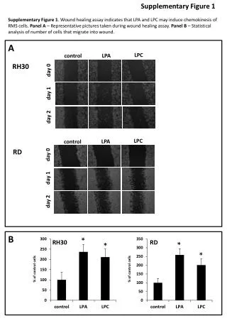
Supplementary Figure 1 presents findings from a wound healing assay demonstrating that lysophosphatidic acid (LPA) and lysophosphatidylcholine (LPC) significantly enhance the chemokinetic migration of RMS cells. Panel A features representative images from the assay, while Panel B provides statistical analysis of the cell migration quantified at day 0, 1, and 2 for control, LPA, and LPC conditions in RH30 and RD cell lines. The results show a marked increase in the percentage of cells migrating into the wound area compared to control.

Wound Healing Assay Reveals Induction of Chemokinesis by LPA and LPC in RMS Cells
E N D
Presentation Transcript
Supplementary Figure 1 Supplementary Figure 1. Wound healing assay indicates that LPA and LPC may induce chemokinesis of RMS cells. Panel A – Representative pictures taken during wound healing assay. Panel B – Statistical analysis of number of cells that migrate into wound. A LPC LPA control RH30 day 0 day 1 day 2 LPC LPA control RD day 0 day 1 day 2 B * RH30 RD * * * % of control cells % of control cells