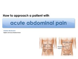
Acute Limb Pain
Acute Limb Pain. Myra Lalas Pitt Morning Report 9/12/11. Differential Diagnosis. Orthopedic/Mechanical Slipped capital femoral epiphysis Legg-Calvé-Perthes disease Trauma/Overuse Fracture Soft-tissue injury Osgood-Schlatter disease Hypermobility. Infection/Infection-related

Acute Limb Pain
E N D
Presentation Transcript
Acute Limb Pain Myra Lalas Pitt Morning Report 9/12/11
Differential Diagnosis • Orthopedic/Mechanical Slipped capital femoral epiphysis Legg-Calvé-Perthes disease • Trauma/Overuse Fracture Soft-tissue injury Osgood-Schlatter disease Hypermobility • Infection/Infection-related Septic arthritis Osteomyelitis Reactive arthritis Rheumatic fever Lyme disease Toxic synovitis
Differential Diagnosis • Inflammatory Juvenile idiopathic arthritis Systemic lupus erythematosus Henoch Schönlein purpura • Noninflammatory Growing pains Fibromyalgia Conversion reaction • Malignancy Leukemia Neuroblastoma Bone tumors • Hematologic Hemophilia Sickle cell anemia
Slipped Capital Femoral Epiphysis • Noninflammatory condition in which the femoral head is displaced from the femoral neck • Commonly affects overweight boys between 10-14 yo • can be associated with endocrine disorders such as hypothyroidism or pituitary deficiencies (eg, growth hormone deficiency)
The left panel demonstrates displacement of the femoral head from the femoral neck in the left hip. Orthopedic correction includes realignment and surgical fixation with a central screw and is depicted in the right panel. Tse S M L , Laxer R M Pediatrics in Review 2006;27:170-180
History: may report a preceding history of trauma and often presents with pain and an inability to walk • PE: may show a limb held slightly flexed and externally rotated. Passive internal rotation of the hip often is limited and painful. • Treatment: no weight bearing until seen by Ortho; surgical fixation done • Prognosis: good but at risk for avascular necrosis of the hip • Follow up: do close follow up because the contralateral hip can be involved in up to 1/3 of cases.
Legg-Calve-Perthes Disease • Avascular necrosis of the capital femoral epiphysis • Theoretical cause: repeated interruptions of the vascular supply to the femoral heads. • Commonly occurs in boys between 4-10 yo. • Presents with a limp, pain, and reduced hip ROM • Treatment: maintaining the femoral head within the acetabulum by abduction splints or casts or surgically with an osteotomy of the proximal femur.
Tse S M L , Laxer R M Pediatrics in Review 2006;27:170-180 Radiographs of various stages of Legg-Calvé-Perthes disease. Progressive changes of the left proximal femur include Stage 1: initial joint space widening and irregularity of the physis, Stage 2: fragmentation, Stage 3: reossification, and Stage 4: healing.
Osgood Schlatter Disease • Osteochondritis of the tibial tubercle • Traction apophysitis of the proximal tibial tubercle at the insertion of the patellar tendon • Characterized by pain and swelling at the tibial tubercle, the point of insertion of the patellar tendon
Presentation: usually 13-14 yo males; anterior knee pain that worsens over time • Diagnosis: by PE Xrays are not necessary unless the patient has atypical complaints (pain that awakens the patient at night, pain at rest, pain not directly over the tibial tubercle, associated systemic complaints) • Management: analgesics, wearing a protective pad over the tibial tubercle, PT; can participate in sports
Transient Synovitis Most common cause of hip pain in childhood. Self-limited inflammatory condition caused by a nonpyogenic inflammatory response of the synovium Peak incidence: 3-6 yo M > F, has a slight predilection for the right side Presentation: hip or groin pain is the most common initial symptom, but referred pain to the medial aspect of the thigh or knee is found in 10% to 30% of patients. Affected patients either walk with a limp or, with severe pain, refuse to walk at all. The leg is held in flexion with slight abduction and external rotation.
PE: passive movement is usually pain-free; however, there may be pain and a slightly decreased range of motion with extreme internal rotation or abduction. • Diagnosis: One of exclusion Good H & P CBC, ESR, CRP AP and "frog-leg" lateral views of the pelvis
Differentiating Septic Arthritis from Transient Synovitis (from Texas Children’s Hospital Handbook)
Linear periosteal reaction is extensive around the distal femoral metaphysis. Bone destruction is noted around the distal femoral metaphysis posteromedially (arrow). Tse S M L , Laxer R M Pediatrics in Review 2006;27:170-180
Growing Pains • Occur in children between 3-12 yo • Characterized by intermittent nighttime nonarticular pain most commonly in the legs; typically bilateral; not associated with limping • PE: normal • Treatment: heat, massage, and analgesics; Reassurance
References Kimura Yukiko, "Chapter 207. Musculoskeletal Pain Syndromes" (Chapter). Colin D. Rudolph, Abraham M. Rudolph, George E. Lister, Lewis R. First, Anne A. Gershon: Rudolph's Pediatrics, 22e: http://www.accesspediatrics.com/content/7019794. Lowry AW, Bhakta KY, Nag PK, "Chapter 13. Emergency Medicine" (Chapter). Lowry AW, Bhakta KY, Nag PK: Texas Children's Hospital Handbook of Pediatrics and Neonatology:http://www.accesspediatrics.com/content/7436297. McQuillen Kemedy K, "Chapter 105. Inflammatory Musculoskeletal Disorders" (Chapter). Gary R. Strange, William R. Ahrens, Robert W. Schafermeyer, Robert A. Wiebe: Pediatric Emergency Medicine, 3e: http://www.accesspediatrics.com/content/5341179. Tse, S. and R. Laxer. Approach to Acute Limb Pain in Childhood. Pediatrics in Review Vol. 27 No. 5 May 1, 2006 pp. 170 -180 www.uptodate.com
PREP Questions A 12-yo boy presents with an itchy rash that you diagnose as scabies. As he leaves the exam room, you note that he is limping. He is overweight, and his mother states that he has been playing football to get some exercise. She believes he is limping because he was injured during football practice several weeks ago & has been complaining of L knee pain. Findings on PE of the knee are normal, but he complains of pain with hip motion.
Of the following, the radiographic study most likely to yield a diagnosis is • AP, lateral, & sunrise radiographs of the knee • Bilateral AP & frog leg radiographs of the hips • MRI of the knee • Ultrasound of the hip • Ultrasound of the knee