Lipids (Lipid Profile)
Lipids (Lipid Profile). Lab. 8. Introduction. The major lipids present in the plasma are: fatty acids, Triglycerides, cholesterol and phospholipids. Other lipid-soluble substances, present in much smaller amounts (e.g. steroid hormones ).

Lipids (Lipid Profile)
E N D
Presentation Transcript
Lipids(Lipid Profile) Lab. 8
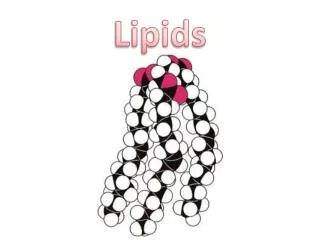
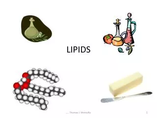
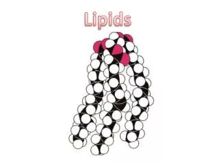
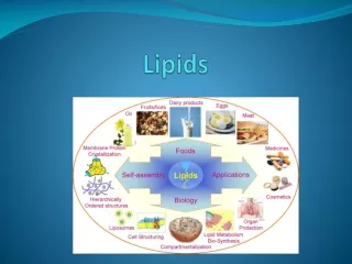
Introduction • The major lipids present in the plasma are: • fatty acids, • Triglycerides, • cholesterol • and phospholipids. • Other lipid-soluble substances, present in much smaller amounts (e.g. steroid hormones ). • Elevated plasma concentrations of lipids, particularly cholesterol, are related to the pathogenesis of atherosclerosis.
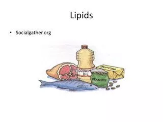
Lipids transport • Lipids are carried in the bloodstream by complexes known as lipoproteins. • This is because these lipids are not soluble in the plasma water. • Thus they travel in micelle-like complexes composed of phospholipids, cholesterol and protein on the outside with cholesteryl esters, and triglycerides on the inside. • The four main types of lipoproteins are chylomicrons, VLDL, LDL, and HDL
Clinical Significance • Cholesterol and triglycerides, like many other essential components of the body, attract clinical attention when present in abnormal concentrations. • Increased or decreased levels usually occur because of abnormalities in the synthesis, degradation, and transport of their associated lipoprotein particles. • Increased or decreased plasma lipoproteins are named hyperlipoproteinemia & hypolipoproteinemia respectively.
Lipids as Biochemical Markers of Disease • Clinical chemistry laboratories offer many tests for lipid disorders. • One of the most common tests is the lipid profile. • This panel of tests includes measurement of triglycerides and cholesterol in the form of lipoprotein-cholesterol molecules, • low density lipoprotein cholesterol (LDL-C) • and high-density lipoprotein cholesterol (HDL-C). • The results of testing for these lipids provide measures of risk for coronary artery disease.
It is therefore clear that lipid measurements should be made in all patients known to have vascular disease, and in those at increased risk. • Thus plasma lipids should be measured in the individuals with the following: • CHD (and cerebrovascular and peripheral vascular disease) • a family history of premature coronary disease (occurring at age <60 years) • other major risk factors for CHD (e.g. diabetes mellitus, hypertension) • patients with clinical features of hyperlipidaemia • patients whose plasma is seen to be lipaemic.
Triglycerides Glycerol Triglyceride • Glycerol backbone with FA attached by ester bonds • Sources of Triglycerides: • Exogenous source: Dietary • Endogenous : Liver and tissue storage
Triglycerides • Serum triglycerides measurements are done for the following clinical reasons: • Hypertriglyceridemiaincreases the risk for pancreatitis. • Hypertriglyceridemiais associated with the following clinical findings: eruptive xanthoma, lipemiaretinalis, hepatomegaly, splenomegaly, depressed HDL-cholesterol. • in characterizing risk of CVDs • For the estimation of LDL-cholesterol, using the Friedewald equation
Specimen • Serum, Plasma (EDTA) • Certain anticoagulants, such as fluoride, citrate, and oxalate, cause large shifts of water from the red blood cells to the plasma, which result in the dilution of plasma components ( as much as 10%) • Fasting sample (from 12 to 16 h) is essential for triglyceride analysis • Samples drawn from nonfasting patients are not suitable for analysis, since elevated TG levels caused by normal absorption of food cannot be distinguished from elevated TG resulting from abnormal lipid metabolism or inborn errors of metabolism. • Storage and stability
Enzymatic Method • Glycerol, released from triglycerides after hydrolysis with lipoprotein lipase, • Tranformed by glycerolkinase into glycerol-3-phosphate • which is oxidized by glycerolphosphate oxidase into dihydroxyacetone phosphate and hydrogen peroxide. • In the presence of peroxidase, the hydrogenperoxide oxidizes the chromogen 4-aminophenazone/ESPT to form purplequinoneimine whose intensity is proportional to theconcentration of triglycerides in the sample.
Enzymatic Method Triglycerides Glycerol + 3 fatty acids Glycerol + ATP Glycerol-3 phosphate + ADP Glycerol-3 phosphate dihydroxyacetone + H2O2phosphate H2O2 + 4-aminophenazone/ESPT Quinoneimine Lipoprotein lipase glycerolkinase glycerolphosphate oxidase peroxidase
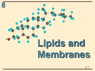
Cholesterol • Cholesterol is a sterol compound that is found in all animal tissues • Serves many important physiological functions including: • synthesis of bile acids, • steroid hormones, • and cell membranes. • Cholesterol also appears to be involved in atherosclerosis; thus cholesterol measurement is one of the most common laboratory tests used today.
Specimen • Serum, Plasma (EDTA, Heparin) • Certain anticoagulants, such as fluoride, citrate, and oxalate, cause large shifts of water from the red blood cells to the plasma, which result in the dilution of plasma components. • Storage and Stability • 7 days at 20 – 25 °C • 7 days at 4 – 8 °C • 3 months at -20 °C
Enzymatic Reaction • Determination of cholesterol after enzymatic hydrolysis and oxidation. • The colorimetric indicator is quinoneimine which is generated from 4-aminoantipyrine and hydroxybenzoate by hydrogen peroxide under the catalytic action of peroxidase Cholesterol Esterase Cholesterol oxidase Cholesterol-3-one Peroxidase
HDL • HDL is a fraction of plasma lipoproteins • It is composed of: • 50% protein, • 25% phospholipid, • 20% cholesterol, • and 5% triglycerides • Evidence suggests that high-density lipoprotein (HDL) cholesterol is a primary coronary heart disease (CHD) risk factor.
Analysis Methods • Ultracentrifugation • Polyacrylamide Gel Electrophoresis • Precipitation • based on the ability of various agents to precipitate selectively the major lipoprotein fractions, except HDL • Immunological • Antibodies against human lipoproteins are used to form antigen-antibody complexes with LDL, VLDL and chylomicrons in a way that only HDL-cholesterol is selectively determined by an enzymatic cholesterol measurement
Precipitation Method • In the plasma, cholesterol is transported by three lipoproteins: high density lipoprotein, low density lipoprotein, and very low density lipoprotein • HDL lipoproteins are assayed, after precipitation of LDL and VLDL lipoproteins with polyethylene glycol (PEG) 6000. • HDL is left in the supernatant solution for cholesterol quantitation.
Low Density Lipoprotein [LDL-chol] = [Total chol] - [HDL-chol] - ([TG]/2.2) • Where all concentrations are given in mmol/L • (note that if calculated using all concentrations in mg/dL then the equation is: [LDL-chol] = [Total chol] - [HDL-chol] - ([TG]/5) • The quotient ([TG]/5) is used as an estimate of VLDL-cholesterol concentration. • It assumes, first, that virtually all of the plasma TG is carried on VLDL, and second, that the TG:cholesterol ratio of VLDL is constant at about 5:1
Limitations of the Friedewald equation • The Friedewald equation should not be used under the following circumstances: • when chylomicrons are present • when plasma triglyceride concentration exceeds 400 mg/dL • in patients with dysbetalipoproteinemia (type III hyperlipoproteinemia