Download
1 / 41
450 likes | 1.01k Vues
This comprehensive guide outlines regulatory guidelines and best practices for setting up a molecular laboratory to prevent contamination. It covers spatial separation, equipment setup, ventilation systems, and disinfection protocols. The article details contamination sources and prevention methods, including unidirectional flow and mechanical barriers. Examples of laboratory design and operations are provided, emphasizing the importance of maintaining a clean and organized workspace.

E N D
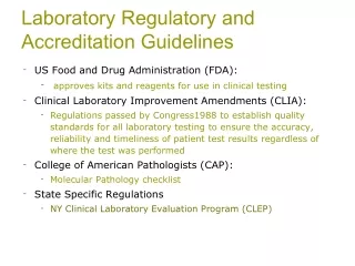
Laboratory Regulatory and Accreditation Guidelines • US Food and Drug Administration (FDA): • approves kits and reagents for use in clinical testing • Clinical Laboratory Improvement Amendments (CLIA): • Regulations passed by Congress1988 to establish quality standards for all laboratory testing to ensure the accuracy, reliability and timeliness of patient test results regardless of where the test was performed • College of American Pathologists (CAP): • Molecular Pathology checklist • State Specific Regulations • NY Clinical Laboratory Evaluation Program (CLEP)
Professional Guidelines • American College of Medical Genetics (ACMG) • Standards and Guidelines for Clinical Genetics Laboratories • Clinical and Laboratory Standards Institute (CLSI) • MM01-A2: Molecular Diagnostic Methods for Genetic Diseases • MM05-A2: Nucleic acid amplification assays for molecular hemathpathology • MM09-A: Nucleic acid sequencing methods in diagnostic laboratory medicine • MM13-A: Collection, Transport, Preparation, and Storage of Specimens for Molecular Methods • MM14-A: Proficiency Testing (External Quality Assessment) for Molecular Methods • MM17-A: Verification and Validation of Multiplex Nucleic Acid Assays • MM19-P: Establishing Molecular Testing in Clinical Lab Environments • MM20-A: Quality Management for Molecular Genetic Testing • NBS06-A: Newborn Blood Spot Screening for Severe Combined Immunodeficiency by Measurement of T-cell Receptor Excision Circles
Contamination Introduction of unwanted nucleic acids into specimen - the sensitivity of PCR techniques makes them vulnerable to contamination Repeated amplification of the same target sequence leads to accumulation of amplification products in the laboratory environment • A typical PCR generates as many as 109 copies of target sequence • Aerosols from pipettes will contain as many as 106 amplification products • Buildup of aerosolized amplification products will contaminate laboratory reagents, equipment, and ventilation systems
Potential Sources of Contamination Cross contamination between specimens Amplification product contamination Laboratory surfaces Ventilation ducts Reagents/supplies Hair, skin, saliva, and clothes of lab personnel
Setting Up a Molecular Laboratory • Mechanical barriers to prevent contamination • Spatial separation of pre- and post-amplification work areas • Area 1 – Reagent preparation • Area 2 – Specimen preparation, PCR set-up • Area 3 – Amplification/product detection, plasmid preparation • Physically separated and, preferably, at a substantial distance from each other
Unidirectional Flow Both personnel and specimens Amplification product-free to product-rich Remove PPE before leaving one area Avoid or limit reverse direction Reusable supplies in the reverse direction need to be bleached.
Features of the 3 Areas • Each area has separate sets of equipment and supplies • Refrigerator/freezer (manual defrost) • Pipettes, tips, tubes, and racks • Centrifuge, timers, vortex • Lab coat (color-coded), disposable gloves, safety glasses, and other PPE • Cleaning supplies • Office supplies • Ventilation system • Dead air box with UV light – serves as a clean bench area
Features of the 3 Areas • Air pressure • Reagent Prep and Specimen Prep – Positive • Postamplification - Negative • Reagent Prep – Single entrance, reagents used for amplification should not be exposed to other areas • Specimen Prep – Specimens should not be exposed to post-amplification work areas
Laboratory Design ExampleMitchell P. S. et al. Nucleic Acid Amplification Methods: Laboratory Design and Operations, 2004, In “Molecular Microbiology: Diagnostic Principles and Practice, edited by D. H. Persing et al” 99. 85-93.
Two Areas Only • Area 1 – Reagent prep, specimen prep, and target loading – use of laminar-flow hoods • Area 2 – Amplification/product detection
Alternative to Spatial Separation Class II biological safety cabinet Dedicated areas for each work phase Unidirectional Automated specimen processing station/closed-tube amplification and detection system
Chemical and Enzymatic Barriers • Work stations should all be cleaned with 10% sodium hypochlorite solution (bleach), followed by removal of the bleach with ethanol. • Ultra-violet light irradiation • UV light induces thymidine dimers and other modifications that render nucleic acid inactive as a template for amplification • Enzymatic inactivation with uracil-N-glycosylase • Substitution of uracil (dUTP) for thymine (dTTP) during PCR amplification • New PCR sample reactions pre-treated with Uracil-N-glycosylase (UNG) – contaminating PCR amplicons are degraded leaving only genomic DNA available for PCR
Important Details • Use of positive displacement pipettes and disposable filtertip pipette tips • Avoid production of aerosols when pipetting • Use of sterilized single-use plasticware • Use of cleanroom floor mats • Minimizes the risk of amplicon carry-over on clothing, hair and skin • Hairnet • Dedicated safety glasses • Disposable labcoat/gown • Gloves • Shoe covers
More Important Details • Use of nuclease free or autoclaved water • Aliquot oligonucleotides – multiple freeze thaws will cause degradation • Always include a blank (no template) control to check for contamination • Wipe test • Monthly • Detect and localize the contamination • Identify the source of the contamination
Decontamination Approaches • Clean the work area & equipments routinely • Clean the PCR workstation at the start and end of each work day/run (UV light, 70% ethanol, fresh 10% sodium hypochlorite, DNA Away) • Clean the exterior and interior parts of the pipette • Clean the equipment • Clean the doorknobs, handle of freezers
Other Considerations • Temperature and humidity requirements • Exhaust ventilation • Water quality • Back-up power system • Eye wash
When is a Validation/Verification Study Required? • Introduce a new testing system • New analyte • Analyte previously measured/detected on an alternate system • An analyte added to a test system • A modification to a test system • Applies to • Unmodified, FDA-cleared or approved method • Modified, FDA-cleared or approved method • In-house method • Standardize method such as textbook procedure • Determine analytic performance of an assay
Assay Validation • Accuracy: Verify the method produces the correct results • Test reference materials (known positive and negative specimens) • Compare test results vs. reference method • Compare split sample results • Compare results to clinical diagnosis • Sample for accuracy study • Patient samples with known results • QC materials • PT materials
Assay Validation (cont.) • Precision: Measure of the reproducibility • Day-to-day variance • Run-to-run variance • Within-a-run variance • Operator variance • Repeat testing of samples, e.g. known patient or QC samples, over time
Assay Validation (cont.) • Analytical Sensitivity: Minimum detection limit • Quantitate amount of RNA or DNA extracted • Control material of known concentration or copy number • LOD, LOQ, LOB
Assay Validation (cont.) • Analytical Specificity: Detect only the analyte intended to be measured • Interfering Substances: Document from product information, literature, or own testing • Anticoagulant • Specimen type (DBS) • Reportable Range: Upper and lower limits of the testing system, presence and absence of mutations • Reference Interval: Document the normal values • Carryover study • Stability Study
Conducting a Validation Study • Planning • Determine the number and type of specimens • Study duration • Establish acceptance criteria • Method limitation • State the methods to resolve discrepancies • Testing • Data Collection and Analysis • Resolving Discrepancies • Sequence amplicon • Test sample by another laboratory
Conducting a Validation Study (cont.) • Implementation • Review and Approval by Lab Director • SOP • Assure ongoing QA • PT
Reagents • Labeling Reagents: • Content, quantity, concentration • Lot # • Storage requirements (temperature etc.) • Expiration date • Date of use/disposal • Know your critical reagents (enzymes, probes, digestion and electrophoresis buffers) and perform QC checks as appropriate
Critical Molecular Assay Components • Nucleic Acids: Prepare aliquots appropriate to workflow to limit freeze-thaw cycles • Primers and probes • dNTPs • Genomic DNA • 4-8°C • -15 to -25°C • Enzymes • Benchtop coolers recommended • Fluorescent reporters • Limit exposure to light • Amber storage tubes or wrap in shielding (foil)
Controls for Each Run Appropriate positive, negative and no template controls (extraction blank) should be included for each run of specimens being tested
Molecular Assay Controls • Positive and negative controls: • Inhibitors • Component failure • Interpretation of results • Sources: • Residual positive DBS • PT samples • QC materials through purchase or exchange • No template controls: • Nucleic acid contamination
Positive Controls Ideally should represent each target allele used in each run May not be feasible when: • Highly multiplex genotypes possible • Systematic rotation of different alleles as positives • Rare alleles • Heterozygous or compound heterozygous specimens
Positive Controls • Assays based on presence or absence of product • Internal positive amplification controls to distinguish true negative from false due to failure of DNA extraction or PCR amplification • PCR amplification product of varying length • Specimens representing short and long amplification products to control for differential amplification • Quantitative PCR • Controls should represent more than one concentration • Control copy levels should be set to analytic cut-offs
In Newborn Screening How can you control for presence of sufficient amount/quality of DNA for a PCR based test in a NBS lab?
PCR with Internal Controls Tetra-primer ARMS-PCR Simultaneous amplification of: Positive amplification control Mutation allele Reference allele Alternative to tetra-primer ARMS is to include an additional primer set to amplify a different control sequence
False Negative: ADO Allele drop-out (ADO): the failure of a molecular test to amplify or detect one or more alleles • Potential causes: • DNA template concentration • Incomplete cell lysis • DNA degradation • Non-optimized assay conditions • Unknown polymorphisms in target sites • Reagent component failure • Major concern for screening laboratories • Confirmation of mutation inheritance in families may not an option
False Positives • Potential causes: • Non-optimized assay conditions • Unknown polymorphisms in target sites • Gene duplications • Oligonucleotide mis-priming at related sequences • Psuedogenes or gene families • Oligonucleotide concentrations too high • Nucleic acid cross-contamination
Sample Acceptance and Tracking • Special specimen acceptance criteria? • Assign a unique code to each patient • Use two patient-identifiers at every step of the procedure • Develop worksheets and document every step • Positive ID
Proficiency Testing • Assessment of the Competence in Testing • Required for all CLIA/CAP certified laboratories • Performed twice a year • If specimens are not commercially available alternative proficiency testing program has to be established (specimen exchange etc.)
Molecular Assay Proficiency Testing Material Sources • CDC NSQAP • UKNEQS • EuroGentest • CAP • Maine Molecular • SeraCare • Corielle • ECACC • In-house samples • Round-robin with other NBS laboratories
Mutation Nomenclature • Uniform mutation nomenclature • Den Dunnen & Antonarakis (2001) Hum Genet 109:121-124 • Den Dunnen & Paalman (2003) Hum Mutat 22:181-82 • Human Genome Variation Society (http://www.hgvs.org/mutnomen/) • Conventional notation should be retained for “established” clinical alleles
STANDARD NOMENCLATURE FOR GENES AND MUTATIONS Nucleotide numbering based on a coding DNA sequence Standard mutation nomenclature based on a coding DNA sequence Source: Ogino, et al (2007) J Mol Diagn 9:1-6
Examples of Mutation Nomenclature: CFTR *Conventional CFTR exon/intron numbering includes exons 6a and 6b, exons 14a and 14b, and exons 17a and 17b; for exon/intron numbers in parentheses, these exon pairs are numbered sequentially without modifiers such as ′6a′ and ′6b.′
Other QA/QC Considerations • Laboratory Cleanliness and Waste Disposal • Instrument Maintenance and Calibration • Instrument/Method Comparison • Document Management • Turnaround Time or Other QA Monitors • Personnel Training and Competency • Periodic Review of QA/QC • COOP Plan