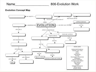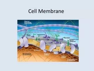
Cell Biology 4 - Cell membrane
310 likes | 481 Vues
Cell Biology 4 - Cell membrane. Sung Youn Lee, PhD. Student Veterinary collage, Room 320 02 450 3719, 016 293 6059 leevet@paran.com. Clinical Case. Patient: Cat, domestic shorthair, 3 years old Presenting Signs and Complaints: Gone for 10 days, returned showing labored breathing

Cell Biology 4 - Cell membrane
E N D
Presentation Transcript
Cell Biology 4- Cell membrane Sung Youn Lee, PhD. Student Veterinary collage, Room 320 02 450 3719, 016 293 6059 leevet@paran.com
Clinical Case • Patient: Cat, domestic shorthair, 3 years old • Presenting Signs and Complaints: Gone for 10 days, returned showing labored breathing • Shallow rapid respirations: no lung sounds ausculated; dull chest on percussion • Radiograph: Bilateral fluid in chest • Problem List: 1. Fluid in chest. 2. Dyspnea
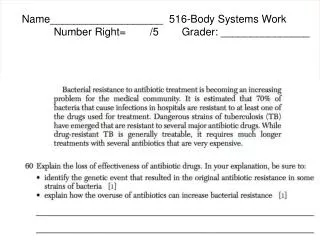
The expected response was segmented/band neutrophil with toxic granulation. Toxic granulation may be defined as dark, basophilic granulation in the cytoplasm which usually varies from fine to coarse in consistency and from few to numerous in number.It is thought to be the toxic precipitation of RNA and is a stress response to infection or inflammation.
Hemogram • There is normocytic normochromic nonregenerative anemia. In this case anemia may be associated with feline leukemia virus(FeLV) infection or the anemia of chronic disease. • On first appearance leukopenia and mature neutropenia suggest a peracute inflammatory response, perhaps related to pyrothorax. However, the absence of either a degenerative a regenrative left shift or a mature neutrophilia in relation to the pyothorax leads one to suspect an underlying bone marrow problem affecting myelopoiesis.
Hemogram • The FeLV infection would be the most likely candidate to cause such a bone marrow disturbance. The toxic morphology of the neutrophil is compatible with a bacterial infection. These morphologic changes occur in the bone marrow, and, although FeLV can cause morphologic aberrations, these finding are most suggestive of an underlying bacterial infetion. • These is a thrombocytopenia that may be associated with the bone marrow abnormalities although an endotoxin-induced thrombocytopenia cannot be ruled out. • Hyperfibrinogenemia is compatible with the inflammatory process.
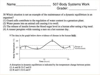
Bone marrow examination • Prior to the initiation of medical management of the pyrothorax a bone marrow examination was completed. The specimen was 80% celluar, the M:E ratio was 2.7:1.0, megakaryocytes were sparse and had abnormal morphology. The erythroid series was present and complete through the metarubric stage with few polychromatophils. The myeloid series was present and complete to the myelocyte stage, but band and segmented neutrophils were rare.
Diagnosis • FeLV infection producing bone marrow dysplasia. Pyrothorax.
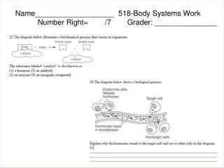
An occasional cat with FeLV will have uneven pupils, called anisocoria
ELISA kit FeLV+ FeLV+ & FIV+ FIV+
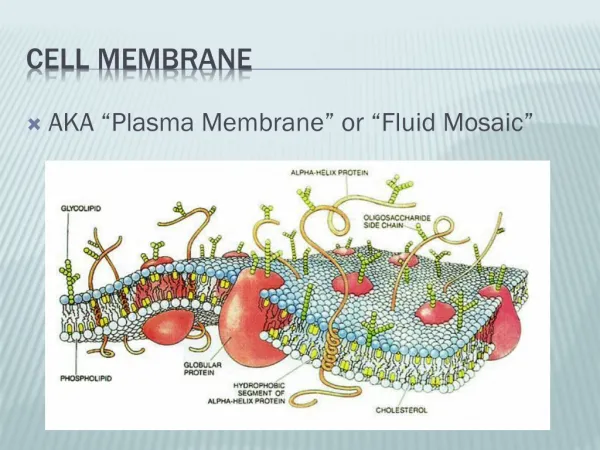
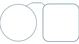
History • 1925 : Gorder and Grendel - bilayer • 1930 : Danielli and Davson –contain protein • 1950 : Danielli –protein_transport polar molecules • 1972 : fluid mosaic model • ~now : peripheral proteins are also functional and not all membrane proteins are freely mobile.
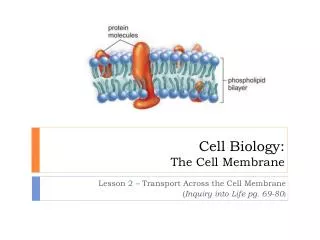
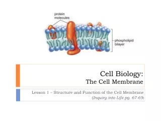
Lipid composition of biological membrane • 40~80% of membrane • Phospholipids, cholesterols, glycolipids
Phospholipids • most prevalent lipids (50% by weight) • 50:1 = phospholipids:protein • 4 major phopholipids • PC (phosphatidylcholine) • PS (phosphatidylserine) • PE (phosphatidylethanolamine) • PI (phosphatidylinositol) • Spingomyelin
cis-double bond Membrane lipids: Glycerophospholipids Phosphatidylcholine Polar Non-polar Alberts Fig. 10-2
Membrane lipids: Glycosphingolipids Polar HO Non-polar (ceramide)
Cholesterols • Amphipathic (polar –OH, hydrophobic –ring) Chelesterol Phospholipid Polar -OH Head Hydrophobic Ring Tail
choline Hydrophobic tails Membrane lipids: Sphingomyelin phosphate Phosphatidylcholine Sphingomyelin Alberts Fig. 10-2 Devlin Fig. 12-13
Glycolipids • The sugar residues of plasma membrane glycolipids almost always face the outside of the cell, that is, they have an asymmetric distribution. • Cell-cell and cell-interstitial matrix interaction.
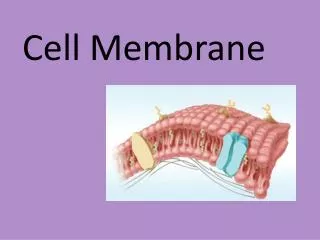
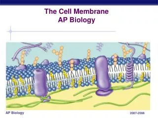
A PLASMA MEMBRANE Integral protein Devlin Fig. 12-22
Membrane structure • Whereas membrane lipids form the foundation of the bilayer, membrane proteins are primarily responsible for function. • Transport of ions and polar molecules, binding of hormones, signal transduction across the membrane, and structure stabilization of the bilayer. • Protein:Lipid ration various • Myelin 0.23 • Mitochondria inner membrane 3.2
Biological membranes: • Always have some protein • Different levels of lipids and proteins