Download
1 / 1
10 likes | 82 Vues
INTRODUCTION. RESULTS. Lack of hippocampal VEGF and VEGFR2 expression in the hypoxia tolerant naked mole rat Dan McCloskey 1 , Sharry Goldman 2 , Bruce Goldman 2 1 Psychology and Neuroscience, College of Staten Island/CUNY; 2 Ecology and Evolutionary Biology, Univ. of Connecticut, Storrs.

E N D
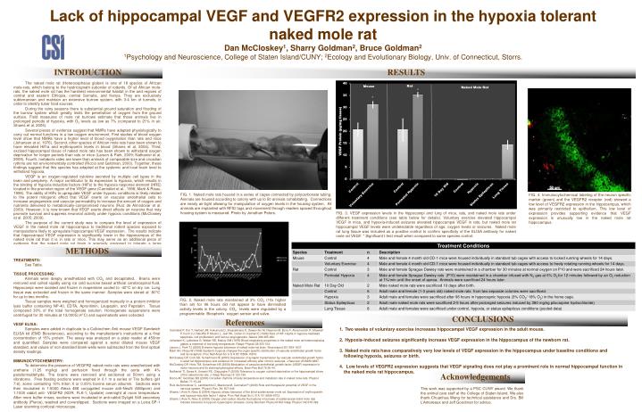
INTRODUCTION RESULTS Lack of hippocampal VEGF and VEGFR2 expression in the hypoxia tolerant naked mole rat Dan McCloskey1, Sharry Goldman2, Bruce Goldman21Psychology and Neuroscience, College of Staten Island/CUNY; 2Ecology and Evolutionary Biology, Univ. of Connecticut, Storrs. FIG. 1. Naked mole rats housed in a series of cages connected by polycarbonate tubing. Animals are housed according to colony with up to 50 animals cohabitating. Connections are nearly air-tight allowing for manipulation of oxygen levels in the housing system. All animals are implanted with RFID tags and movement through readers spaced throughout housing system is measured. Photo by Jonathan Peters. FIG. 4. Immunocytochemical labeling of the neuron specific marker (green) and the VEGFR2 receptor (red) showed a low level of VEGFR2 expression in the hippocampus, which was primarily restricted to epithelium. This low level of expression provides supporting evidence that VEGF expression is unusually low in the naked mole rat hippocampus. FIG. 3. VEGF expression levels in the hippocampi and lung of mice, rats, and naked mole rats under different treatment conditions (see table below for details). Voluntary exercise elevated hippocampal VEGF in mice, and hypoxia-induced seizures elevated hippocampal VEGF in rats, but naked mole rat hippocampal VEGF levels were undetectable regardless of age, oxygen levels or seizures. Naked mole rat lung tissue was included as a positive control to confirm specificity of the ELISA antibody for naked mole rat VEGF. * Significant t-test result when compared to same species control. The naked mole rat (Heterocephalus glaber) is one of 18 species of African mole-rats, which belong to the hystricognath suborder of rodents. Of all African mole-rats, the naked mole rat has the harshest environmental habitat in the arid regions of central and eastern Ethiopia, central Somalia, and Kenya. They are exclusively subterranean and maintain an extensive burrow system, with 3-4 km of tunnels, in order to identify tuber food sources. During the rainy seasons there is substantial ground saturation and flooding of the burrow system which greatly limits the penetration of oxygen from the ground surface. Field measures of mole rat burrows estimate that these animals live in prolonged periods of hypoxia, with O2 levels as low as 7% (compared to 21% in air, Shams et al. 2005). Several pieces of evidence suggest that NMRs have adapted physiologically to carry out normal functions in a low oxygen environment. First studies of blood oxygen level show that NMRs have a higher level of blood oxygenation than rats and mice (Johansen et al. 1976). Second, other species of African mole rats have been shown to have elevated Hif1α and erythropoetin levels in blood (Shams et al. 2004). Third, excised hippocampal tissue of naked mole rats has been shown to withstand oxygen deprivation for longer periods than rats or mice (Larson & Park, 2009; Nathaniel et al. 2009). Fourth, metabolic rates are lower than animals of comparable size and circadian rythms are not environmentally controlled (Riccio and Goldman, 2000). Together, these findings suggest that this species has adapted at the systemic and local brain level to withstand hypoxia. VEGF is an oxygen-regulated cytokine secreted by multiple cell types in the brain and periphery. A major contributor to its expression is hypoxia, which results in the binding of hypoxia-inducible factors (HIFs) to the hypoxia response element (HRE) located in the promoter region of the VEGF gene (Carmeliet et al., 1998; Marti & Risau, 1998). The ability of HIFs to upregulate VEGF under hypoxic conditions is likely related to the potent mitogenic effect that VEGF exerts on vascular endothelial cells to increase angiogenesis and vascular permeability to increase the amount of oxygen and nutrients delivered to metabolically-compromised neurons (Ruiz de Almodovar et al. 2009). However, it is now known that VEGF exerts direct effects on neurons that may promote survival and suppress neuronal activity under hypoxic conditions (McCloskey et al. 2005; 2009). The purpose of the current study was to compare the level of expression of VEGF in the naked mole rat hippocampus to traditional rodent species exposed to manipulations likely to upregulate hippocampal VEGF expression. The results indicate that hippocampal VEGF expression is significantly lower in the hippocampus of the naked mole rat than it is in rats or mice. This may serve as an additional piece of evidence that the naked mole rat brain is specially equipped to tolerate a large variation in oxygen availability. METHODS TREATMENTS: See Table. TISSUE PROCESSING: Animals were deeply anesthetized with CO2 and decapitated. Brains were removed and colled rapidly using ice cold sucrose based artificial cerebrospinal fluid. Hippocampi were isolated and frozen in isopentane cooled to -40°C on dry ice. Lung tissue was extracted and frozen in the same manner. Samples were stored at -80°C for up to two months. Tissue samples were weighed and homogenized manually in a protein inhibitor lysis buffer containing NP-40, EDTA, Apronitinin, Leupeptin, and Pepstatin. Tissue composed 30% of the total homogenate solution. Homogenate suspensions were centrifuged for 30 minutes at 13,000G (4°C) and supernatants were collected. VEGF ELISA: Samples were added in duplicate to a Calbiochem Anti-mouse VEGF Sandwich ELISA kit (EMD Biosciences), according to the manufacturer’s instructions at a final concentration of 15% protein. The assay was analyzed on a plate reader at 450nm and quantified. Samples were compared against a serial diluted mouse VEGF standard, and values of non standard blank wells were subtracted from the final optical density readings. IMMUNOCYTOCHEMISTRY: To determine the presence of VEGFR2 naked mole rats were anesthetized with urethane (1.25 mg/kg) and perfusion fixed through the aorta with 4% paraformaldehyde. The brains were removed and sectioned at 50mm using a vibratome. Free floating sections were washed in 0.1 m a series of Tris buffers (pH 7.6), some containing 10% triton X or 0.05% bovine serum albumin. Sections were then incubated in 1:5000 Alexa 488 conjugated mouse anti-NeuN (Millipore) and 1:1000 rabbit anti- VEGFR2 (KDR, FLK-1; Upstate) overnight at room temperature. After more buffer rinses, sections were incubated in anti-rabbit Dylight 549 secondary antibody (Pierce), washed and coverslipped. Sections were imaged on a Leica SP-1 Laser scanning confocal microscope. FIG. 2. Naked mole rats maintained at 3% CO2 (10x higher than air) for 96 hours did not appear to have diminished activity levels in the colony. CO2 levels were regulated by a programmable Biospherix oxygen sensor and valve. CONCLUSIONS References • Two weeks of voluntary exercise increases hippocampal VEGF expression in the adult mouse. • Hypoxia-induced seizures significantly increase VEGF expression in the hippocampus of the newborn rat. • Naked mole rats have comparatively very low levels of VEGF expression in the hippocampus under baseline conditions and following hypoxia, seizures or birth. • Low levels of VEGFR2 expression suggests that VEGF signaling does not play a prominent role in normal hippocampal function in the naked mole rat hippocampus. Carmeliet P, Dor Y, Herbert JM, Fukumura D, Brusselmans K, Dewerchin M, Neeman M, Bono F, Abramovitch R, Maxwell P, Koch CJ, Ratcliffe P, Moons L, Jain RK, Collen D, Keshert E (1998) Role of HIF-1alpha in hypoxia-mediated apoptosis, cell proliferation and tumour angiogenesis. Nature 394:485-490. Johansen K, Lykkeboe G, Weber RE, Maloiy GM (1976) Blood respiratory properties in the naked mole rat heterocephalus glaber, a mammal of low body temperature. Respir Physiol 28:303-314. Larson J, Park TJ (2009) Extreme hypoxia tolerance of naked mole-rat brain. Neuroreport 20:1634-1637. Marti HH, Risau W (1998) Systemic hypoxia changes the organ-specific distribution of vascular endothelial growth factor and its receptors. Proc Natl Acad Sci U S A 95:15809-15814. McCloskey DP, Croll SD, Scharfman HE (2005) Depression of synaptic transmission by vascular endothelial growth factor in adult rat hippocampus and evidence for increased efficacy after chronic seizures. J Neurosci 25:8889-8897. McCloskey DP, Hintz TM, Scharfman HE (2008) Modulation of vascular endothelial growth factor (VEGF) expression in motor neurons and its electrophysiological effects. Brain Res Bull 76:36-44. Nathaniel TI, Saras A, Umesiri FE, Olajuyigbe F (2009) Tolerance to oxygen nutrient deprivation in the hippocampal slices of the naked mole rats. J Integr Neurosci 8:123-136. Riccio AP, Goldman BD (2000) Circadian rhythms of body temperature and metabolic rate in naked mole-rats. Physiol Behav 71:15–22 Ruiz de Almodovar C, Lambrechts D, Mazzone M, Carmeliet P (2009) Role and therapeutic potential of VEGF in the nervous system. Physiol. Rev. 89: 607-648 Shams I, Avivi A, Nevo E (2004) Hypoxic stress tolerance of the blind subterranean mole rat: Expression of erythropoietin and hypoxia-inducible factor 1 alpha. Proc Natl Acad Sci U S A 101:9698-9703. Shams I, Avivi A, Nevo E (2005) Oxygen and carbon dioxide fluctuations in burrows of subterranean blind mole rats indicate tolerance to hypoxic-hypercapnic stresses. Comp Biochem Physiol A Mol Integr Physiol 142:376-382. Acknowledgements This work was supported by a PSC CUNY award. We thank the animal care staff at the College of Staten Island. We also thank Chuanhua Wang for technical assistance and Drs. Bill L’Amoreaux and Jeff Goodman for advice.