1
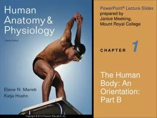
1 . The Human Body: An Orientation: Part B. Anatomical Position. Standard anatomical body position: Body erect Feet slightly apart Palms facing forward. Anatomical Position. Body Planes. Plane: Flat surface along which body or structure is cut for anatomical study. Body Planes.

1
E N D
Presentation Transcript
1 The Human Body: An Orientation: Part B
Anatomical Position • Standard anatomical body position: • Body erect • Feet slightly apart • Palms facing forward
Body Planes • Plane: Flat surface along which body or structure is cut for anatomical study
Body Planes • Sagittal plane • Divides body vertically into right and left parts • Produces a sagittal section • Midsagittal (median) plane • Lies on midline • Parasagittal plane • Not on midline (must add numerical or anatomical qualifier) so many cm from midline; or sternal border, midclavicular, anterior axillary, etc
Body Planes • Frontal (coronal) plane • Divides body vertically into anterior and posterior parts • Transverse (horizontal) plane • Divides body horizontally into superior and inferior parts • Produces a cross section • Oblique section • Cuts made diagonally
Frontal plane Median (midsagittal) plane Transverse plane (a) Frontal section (through torso) (b) Transverse section (through torso, inferior view) (c) Median section (midsagittal) Pancreas Aorta Spleen Liver Spinal cord Intestines Rectum Spleen Left and right lungs Liver Heart Body wall Vertebral column Stomach Arm Subcutaneous fat layer Figure 1.8
Anatomical Variability • Over 90% of all anatomical structures match textbook descriptions, but: • Nerves or blood vessels may be somewhat out of place • Small muscles may be missing
Cephalic Table 1.1
Additional Directional Terms • Ipsilateral – on same side of body • Contralateral – on opposite side of body
Non-synonymous anatomical terms • In a two-legged organism – like the human- superior, cranial, and cephalic are the same • In a two- legged organism inferior and caudal are the same • In a two-legged organism – anterior and frontal are the same and posterior and dorsal are the same • This is not true in a four-legged organism like the dog – different terms become synonymous
Caudal and posterior are the same Dorsum Anterior and Cranial and Cephalic are the same Ventrum
Regional Terms • Two major divisions of body: • Axial • Head, neck, and trunk • Appendicular • Limbs • Regional terms designate specific areas
Body Cavities • Dorsal cavity • Protects nervous system • Two subdivisions: • Cranial cavity • Encases brain • Vertebral cavity • Encases spinal cord
Body Cavities • Ventral cavity • Houses internal organs (viscera) • Two subdivisions (separated by diaphragm): • Thoracic cavity • Abdominopelvic cavity
Cranial cavity Dorsal body cavity Ventral body cavity Cranial cavity (contains brain) Vertebral cavity Superior mediastinum Dorsal body cavity Thoracic cavity (contains heart and lungs) Pleural cavity Pericardial cavity within the mediastinum Vertebral cavity (contains spinal cord) Ventral body cavity (thoracic and abdominopelvic cavities) Diaphragm Abdominal cavity (contains digestive viscera) Abdomino- pelvic cavity Pelvic cavity (contains urinary bladder, reproductive organs, and rectum) (a) Lateral view (b) Anterior view Figure 1.9a-b
Ventral Body Cavities • Thoracic cavity subdivisions: • Two pleural cavities • Each houses a lung • Mediastinum • Contains pericardial cavity • Surrounds thoracic organs • Pericardial cavity • Encloses heart
Ventral Body Cavities • Abdominopelvic cavity subdivisions: • Abdominal cavity • Contains stomach, intestines, spleen, and liver • Pelvic cavity • Contains urinary bladder, reproductive organs, and rectum
Cranial cavity Dorsal body cavity Ventral body cavity Cranial cavity (contains brain) Vertebral cavity Superior mediastinum Dorsal body cavity Thoracic cavity (contains heart and lungs) Pleural cavity Pericardial cavity within the mediastinum Vertebral cavity (contains spinal cord) Ventral body cavity (thoracic and abdominopelvic cavities) Diaphragm Abdominal cavity (contains digestive viscera) Abdomino- pelvic cavity Pelvic cavity (contains urinary bladder, reproductive organs, and rectum) (a) Lateral view (b) Anterior view Figure 1.9a-b
Serous Membrane (Serosa) • Thin, double-layered membrane separated by serous fluid • Parietal serosa lines internal body walls • Visceral serosa covers the internal organs
Outer balloon wall (comparable to parietal serosa) Air (comparable to serous cavity) Inner balloon wall (comparable to visceral serosa) Heart Parietal pericardium Pericardial space with serous fluid Visceral pericardium (b) The serosae associated with the heart. Figure 1.10a-b
Organs in the Thoracic Cavity Figure 22.10a
Abdominopelvic cavity Vertebra Dorsal mesentery Parietal peritoneum Ventral mesentery Visceral peritoneum Peritoneal cavity Alimentary canal organ Liver (a) Schematic cross sections of abdominal cavity illustrate the peritoneums and mesenteries. Figure 23.5a
Abdominopelvic Regions • Nine divisions used primarily by anatomists
Diaphragm Liver Right hypochondriac region Left hypochondriac region Epigastric region Stomach Gallbladder Transverse colon of large intestine Ascending colon of large intestine Right lumbar region Left lumbar region Umbilical region Descending colon of large intestine Small intestine Cecum Initial part of sigmoid colon Right iliac (inguinal) region Hypogastric (pubic) region Left iliac (inguinal) region Appendix Urinary bladder (a) Nine regions delineated by four planes (b) Anterior view of the nine regions showing the superficial organs Figure 1.12
Abdominopelvic Quadrants • Divisions used primarily by medical personnel
Right upper quadrant (RUQ) Left upper quadrant (LUQ) Right lower quadrant (RLQ) Left lower quadrant (LLQ) Figure 1.11
Other Body Cavities • Oral and digestive cavities • Nasal cavity • Orbital cavities • Middle ear cavities • Synovial cavities