Download
1 / 51
530 likes | 654 Vues
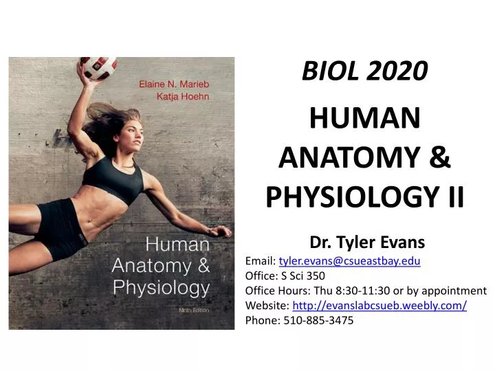
BIOL 2020. HUMAN ANATOMY & PHYSIOLOGY II. Dr. Tyler Evans Email: tyler.evans@csueastbay.edu Office: S Sci 350 Office Hours: Thu 8:30-11:30 or by appointment Website: http ://evanslabcsueb.weebly.com / Phone: 510-885-3475. TODAY’S SCHEDULE. Course Description Cells and Tissues.

E N D
BIOL 2020 HUMAN ANATOMY & PHYSIOLOGY II Dr. Tyler Evans Email: tyler.evans@csueastbay.edu Office: S Sci 350 Office Hours: Thu 8:30-11:30 or by appointment Website: http://evanslabcsueb.weebly.com/ Phone: 510-885-3475
TODAY’S SCHEDULE • Course Description • Cells and Tissues
LEARNING OUTCOMES (What I hope you will gain from this course) • the primary goal of this course is to give you an understanding of anatomy and physiology that will be valuable to you, not only in attaining your career objectives but also in understanding processes that govern your daily life.
LECTURES • lectures will be presented using Powerpoint. • each lecture will be posted on Blackboard prior to class (almost always the evening before). • it is important to realize that my Powerpointslides do not represent all material required for the exams. • Important information that will be covered in exams will be added to each lecture. • thus attending class and taking thorough notes is the key to success.
LECTURES TEXTBOOK AND READINGS • the primary source of information for this course will be the lectures • the textbook will be used mainly to illustrate or clarify materials presented during lectures • however, specific reading assignments may be given throughout the course. • you will be notified of reading assignments and if that reading assignment will be covered on the exam
TEXTBOOK Human Anatomy and Physiology (9th edition) by Elaine N Marieb KatjaHoehn
LABS • you will be performing laboratory exercises designed to help you understand certain fundamental physiological principles. • lab materials will be posted on Blackboard each week • your attendance in lab each week is mandatory • any unexcused absence from a lab will result in 0 credit for the missed lab. • you are required to stay for the entire lab session, or until excused by the instructor. • you will check out with your lab instructor, showing you have met the lab learning objectives and your lab space has been returned to its original condition.
GRADING Your final grade will be determined by three examsand the laboratory component: • exams will focus on lecture materials Midterm Exam #1………..25% (Aug 8) Midterm Exam #2………..25% (Aug 22) Final Exam…………………..25% (Sept 4) Lab Component…...…….25% (Weekly Jul 29-Aug 28) You must pass (>50%) at least one exam to pass the course
COURSE POLICIES (in accordance with CSUEB guidelines) • Academic Dishonesty • please review CSUEB’s policies and understand what is considered academic dishonesty: http://www20.csueastbay.edu/academic/academic-policies/academic-dishonesty.html • Missed Exams • make every effort to avoid missing scheduled exams. In case of an emergency or legitimate conflict, you may be eligible to take a specially scheduled make-up exam. However, you must provide verifiable, written documentation for your absence. Any unexcused absence from an exam will result in a score of 0 for that exam. • Special Academic Accommodations: • if you have a documented disability, accommodations can be arranged for exams and other activities. For more information please visit: http://www20.csueastbay.edu/af/departments/as/ • Courtesy: • Please turn off all audible sounds to any electronic devices (phones, PDAs, etc.) while in lecture and refrain from using your laptops for activities not related to lecture during class time • Use of these items is strictly prohibited during all exams, unless special accommodations have been arranged.
KEYS TO SUCCESS IN BIOL 2020 • this course occurs at twice the speed of a course taken during the regular academic year • for this reason, the time commitment required to receive a good grade in this course is high • DO NOT GET BEHIND: you need to be committed and diligent about reviewing the material covered in lecture • Attend all the lectures • Prepare and take thorough notes • Study those notes • Ask questions! And come see me!
CELLS: THE LIVING UNITS CHAPTER 3 • cells are the structural units of all living things and the smallest living unit • the importance of cells is described by four concepts collectively known as CELL THEORY: A cell is the basic structural and functional unit of living organisms. When you define cell properties, you also define the properties of life The activity of an organism depends on both the individual and the collective activities of its cells The biochemical activities of cells are determined by their form and relative number of subcellular structures Continuity on life from one generation to the next has a cellular basis
CELLS: THE LIVING UNITS CELL STRUCTURE AND FUNCTION • the human body is composed of trillions of cells that vary greatly in shape, size and function. • flat tile-like epithelial cells line the inside of your cheeks and fit tightly together to protect underlying tissues from bacteria • red blood cells are found in the blood and are disc-shaped, allowing these cells to shrink are expand without rupturing • motile cells, such as sperm, have been modified to allow for movement Fig 3.1 pg 62
CELLS: THE LIVING UNITS CELL STRUCTURE AND FUNCTION • regardless of cell type, most cells share the same basic structure • a generalized or COMPOSITE CELL has three main parts: • PLASMA MEMBRANE: the outer boundary of the cell • CYTOPLASM: the intracellular fluid packed with ORGANELLES, small structures that perform specific functions • NUCLEUS: an organelle that coordinates cellular activities. • the nucleus is typically near the cells center Fig 3.2 pg 63
CELLS: THE LIVING UNITS PLASMA MEMBRANE • PLASMA MEMBRANE: the outer boundary of the cell that separates the two major fluid compartments: INTRACELLULAR (inside of cells) and EXTRACELLULAR (outside of cells) • the plasma membrane is not a passive envelope, instead its structure is described by the FLUID MOSAIC MODEL • the membrane is composed of a double layer of lipids (the LIPID BILAYER), in which proteins are inserted • protein components are constantly changing, hence the term fluid mosaic Fig 3.3 pg 64
CELLS: THE LIVING UNITS PLASMA MEMBRANE • membrane proteins make up about 50% of the mass of the plasma membrane and are responsible for most specialized membrane functions • there are two distinct populations of membrane proteins • INTEGRAL PROTEINS: which are firmly inserted into the lipid bilayer • most are TRANSMEMBRANE proteins, meaning they span the entire length of the membrane and protrude on either side • e.g. proteins involved in transport, like ion channels • fig 3.4a pg. 66 • 2. PERIPHERAL PROTEINS: are loosely attached and easily removed without disrupting the membrane • e.g. proteins involved in supporting the cell, such as those of the cytoskeleton • fig 3.4c pg. 66
CELLS: THE LIVING UNITS PLASMA MEMBRANE • membrane proteins play a key role in regulating membrane traffic • our cells are bathed in EXTRACELLULAR FLUID that contains proteins, sugars, ions, fats, etc., and there is constant exchange of these substances across the membrane • substances move through the plasma membrane in two ways: • PASSIVELY: does not require energy • ACTIVELY: does require energy 1. PASSIVE PROCESSES: molecules move from an area where they are in high concentration to areas where they are in lower concentration, that is down or along their concentration gradient e.g. DIFFUSION time time Fig 3.6 pg 68
CELLS: THE LIVING UNITS PLASMA MEMBRANE • 1. PASSIVE PROCESSES • same principle applies to substances moving across cell membranes • DIFFUSION across cell membranes can occur with or without the assistance of membrane proteins Fig 3.7 pg 69
CELLS: THE LIVING UNITS PLASMA MEMBRANE • 2. ACTIVE PROCESSES • refers to the use of energy to move substances across membranes • occurs when substance is not able to dissolve in the lipid bilayer, is too large to pass through channels or must be transported against its concentration gradient • active processes use ATP to change the shape of a transporter protein, such that molecule bound to the transport protein is moved from one side of the membrane to the other. e.g. Sodium-Potassium ATPase (Na+, K+ ATPase) • concentration of K+ is 10X higher in cells than outside and reverse for Na+, • K+ and Na+ slowly leak out of cells by diffusion, so cells actively transport K+ into cells and Na+ out of cells to maintain this gradient
CELLS: THE LIVING UNITS PLASMA MEMBRANE Why would cells invest energy (ATP) to make sure more K+ is inside cells than outside and more Na+outside cells than inside?
CELLS: THE LIVING UNITS CELL JUNCTIONS • though certain cell types occur independently (e.g. blood cells) many other group together into tight communities • three ways in which cells can adhere to one another: • TIGHT JUNCTION: a series of integral proteins in the plasma membranes of adjacent cells fuse together. • e.g. tight junctions in the digestive track prevent digestive enzymes from seeping into the blood stream • DESMOSOME: mechanical couplings that adjoin parts of adjacent cell membranes using proteins called CADHERINS • GAP JUNCTION: a communicating junction between cells. Adjacent cells are connected by hollow tubes CONNEXONS
CELLS: THE LIVING UNITS CELL JUNCTIONS GAP JUNCTION DESMOSOME TIGHT JUNCTION Fig 3.5 pg 67
TISSUE: THE LIVING FABRIC CHAPTER 4 • these cell junctions allows cells to work together to accomplish specific tasks • TISSUES: are groups of cells that are similar in structure and perform a common or related function • four primary tissue types are found throughout the body: • EPITHELIAL (think “covers”) • CONNECTIVE (think “supports”) • NERVOUS (think “controls”) • MUSCLE (think “moves”) • tissues are organized into ORGANS and most organs contain all four tissue types pg 116
TISSUE: THE LIVING FABRIC EPITHELIAL TISSUE • EPITHELIAL TISSUE or an EPITHELIUM is a sheet of cells that covers a body surface or lines a body cavity and forms boundaries between different environments • e.g. form the outer layer of the skin and line the digestive, urogenital and respiratory systems • epithelial tissues have five special characteristics: • 1. POLARITY-epithelium have an APICAL SURFACE that is exposed to the external environment or internal cavity and a BASAL SURFACE facing other cells • because the structure and function of apical and basal surfaces differ, epithelium are said to have APICAL-BASAL POLARITY Fig 4.2 pg 119
TISSUE: THE LIVING FABRIC EPITHELIAL TISSUE • epithelial tissues have five special characteristics: • 2. SPECILAZED CONTACTS: epithelial cells fit tightly together to form a continuous sheet WHAT STRUCTURES WOULD ALLOW CELLS TO BECOME CONNECTED TO FORM TIGHT AND CONTINUOUS SHEETS?
TISSUE: THE LIVING FABRIC EPITHELIAL TISSUE • epithelial tissues have five special characteristics: • 3. SUPPORTED BY CONNECTIVE TISSUE: all epithelial sheets rest upon and are supported by connective tissue • a layer of connective tissue called the BASEMENT MEMBRANE forms along the basal surface of the epithelium • this tissue is rich in the protein COLLAGEN, which helps prevent the epithelium from stretching and tearing Basement membrane
TISSUE: THE LIVING FABRIC EPITHELIAL TISSUE • epithelial tissues have five special characteristics: • 4. AVASCULAR BUT INNERVATED: epithelium contains no blood vessels (i.e. is avascular) but is supplied by nerve fibers (i.e. is innervated) • receive nutrients via diffusion from vessels supplying the basement membrane • 5. REGENERATION: epithelium have a high regenerative capacity and divide rapidly • e.g. skin cells are continually shed and replaced by new ones
TISSUE: THE LIVING FABRIC EPITHELIAL TISSUE • different types of epithelium are classified based on two criteria • the number of cells layers present • The shape of its cells • NUMBER OF CELL LAYERS • SIMPLE EPITHELIA consist of a single cell layer and are typically found where absorption, secretion and filtration occur • STRATIFIED EPITHELIA are composed of two or more layers stacked on top of each other and are common in high abrasion areas like the skin and mouth Fig 4.2a pg 119
TISSUE: THE LIVING FABRIC EPITHELIAL TISSUE • different types of epithelium are classified based on two criteria • the number of cells layers present • the shape of its cells SHAPE OF CELLS • SQUAMOUS CELLS are flattened and scale-like • CUBOIDAL CELLS are box-like • COLUMNAR CELLS are tall and narrow Fig 4.2b pg 119
TISSUE: THE LIVING FABRIC EPITHELIAL TISSUE • different combinations of cell layers and cell shapes give epithelial tissues different properties Fig 4.3a pg 120
TISSUE: THE LIVING FABRIC EPITHELIAL TISSUE • different combinations of cell layers and shape give epithelial tissues different properties Fig 4.3b pg 121
TISSUE: THE LIVING FABRIC EPITHELIAL TISSUE • different combinations of cell layers and shape give epithelial tissues different properties Fig 4.3c pg 121
TISSUE: THE LIVING FABRIC EPITHELIAL TISSUE • different combinations of cell layers and shape give epithelial tissues different properties • nuclei occurring at different heights can give the impression of multiple layers, but is only a single layer (i.e. pseudo-stratified) Fig 4.3d pg 122
TISSUE: THE LIVING FABRIC EPITHELIAL TISSUE • different combinations of cell layers and shape give epithelial tissues different properties • multi-layered: cells divide from below and are pushed to the surface • suitable for protection or areas subject to wear and tear Fig 4.3d pg 123
TISSUE: THE LIVING FABRIC CONNECTIVE TISSUE • most abundant and widely distributed of the primary tissues, but it’s amount in particular organs is variable (skin = high; brain = low) • does more than just connect body parts, also supports (bone/cartilage), protects, insulates, stores reserve energy (fat) and transports substances • like epithelial tissue, connective tissue has distinguishing characteristics: • COMMON ORGIN: all arise from MESENCHYME during embryonic development • DEGREES OF VASCULARITY: dense connective tissue is poorly vascularized, but other types have a rich supply of blood vessels. • EXTRACELLULAR MATRIX: connective tissue is unique in that it is composed largely of non-living tissue called extracellular matrix (ECM) • the ECM gives connective tissue its strength to bear weight and withstand force
TISSUE: THE LIVING FABRIC CONNECTIVE TISSUE • different combinations of cells and fibers (comprised of the proteins COLLAGEN or ELASTIN) give connective tissue different properties AREOLAR CONNECTIVE TISSUE: most common with variety of functions • loosely packed fibers Fig 4.8a pg 131
TISSUE: THE LIVING FABRIC CONNECTIVE TISSUE • different combinations of cells and fibers (comprised of the proteins COLLAGEN or ELASTIN) give connective tissue different properties • ADIPOSE (FAT) TISSUE: similar to areolar, but high nutrient storage capacity with a high quantity of ADIPOCYTES or fat cells, used as energy reserves • also acts as shock absorber and helps the body to retain heat Fig 4.8b pg 131
TISSUE: THE LIVING FABRIC CONNECTIVE TISSUE • different combinations of cells and fibers (comprised of the proteins COLLAGEN or ELASTIN) give connective tissue different properties DENSE REGULAR CONNECTIVE TISSUE: fibers are densely packed and arranged in parallel which gives this tissue great resistance to pulling forces (i.e. HIGH TENSILE STRENGTH) in one direction • forms tendons Fig 4.8d pg 133
TISSUE: THE LIVING FABRIC CONNECTIVE TISSUE • different combinations of cells and fibers (comprised of the proteins COLLAGEN or ELASTIN) give connective tissue different properties ELASTIC CONNECTIVE TISSUE: found in a few ligaments that are very stretchy, such as those connecting the vertebrae, and the walls of large arteries • high concentration of elastin provides “stretchiness” Fig 4.8f pg 134
TISSUE: THE LIVING FABRIC CONNECTIVE TISSUE • CARTILAGE: is intermediate between the dense connective tissue we just described and bone. Cartilage is tough, flexible, avascular and a high water content (reduces compression) • types of cartilage differ by the dominant fiber present: HYALINE CARTILAGE: most abundant with mostly collagen fibers • only a small number of cells called CHONDROCYTES Fig 4.8g pg 135
TISSUE: THE LIVING FABRIC CONNECTIVE TISSUE • BONE (OSSEOUS): is very hard and has an exceptional ability to support the body • similar to the make-up of cartilage, but with additional collagen fibers and CALCIUM SALTS deposited between the fibers • bone cells called OSTEOBLASTS and OSTEOCLASTS secrete these materials Fig 4.8j pg 137
TISSUE: THE LIVING FABRIC MUSCLE TISSUE • MUSCLE TISSUE: highly cellular, well vascularized tissue involved in movement • muscle cells possess MYOFILAMENTS composed of the proteins ACTIN and MYOSIN that function during muscle contraction • there are three types of muscle tissue: • 1.) SKELETAL: attached to the bones of the skeleton and used in movement • possess a striated or “banded” appearance due to the very ordered structure of actin and myosin in muscle cells • 2.) SMOOTH: no striations and found in the walls of hollow organs (e.g. digestive tract and blood vessels) • squeezes substances through these organs by contracting and relaxing • 3.) CARDIAC: found in the walls of the heart and its contraction helps to propel blood through the body • like skeletal muscle, cardiac muscle is striated
TISSUE: THE LIVING FABRIC MUSCLE TISSUE • 1.) SKELETAL: attached to the bones of the skeleton and used in movement • possess a striated or “banded” appearance due to the very ordered structure of actin and myosin in muscle cells Fig 4.9a pg 138
TISSUE: THE LIVING FABRIC NERVOUS TISSUE NERVOUS TISSUE: main component of brain, spinal cord and nerves which regulate and control body functions Composed of two main cells: 1. NEURONS: cells specialized for transmitting electrical signals and therefore possess long thin extensions 2. SUPPORTING CELLS: non-conductive and perform a number of tasks Fig 4.10 pg 140
GLOSSARY OF TISSUE HISTOLOGY • histology and tissue types covered in previous slides may be on lecture exams • the lab component of this course covers many more tissue types than presented in today’s lecture • for this reason I have included extra slides with additional tissue histology • you will not need to know these slides for lecture exams TRANSITIONAL EPITHELIUM Fig 4.3d pg 123
GLOSSARY OF TISSUE HISTOLOGY RETICLUAR CONNECTIVE TISSUE • fibers are softer and this type of connective tissue is more delicate Fig 4.8c pg 132
GLOSSARY OF TISSUE HISTOLOGY DENSE IRREGULAR CONNECTIVE TISSE • collagen fibers are thicker and arranged in different directions, so can withstand force from many different directions Fig 4.8e pg 133
GLOSSARY OF TISSUE HISTOLOGY • CARTILAGE: is intermediate between the dense connective tissue we just described and bone. Cartilage is tough, flexible, avascular and a high water content (reduces compression) • types of cartilage differ by the dominant fiber present: ELASTIC CARTILAGE more elastin fibers so much more stretchable Fig 4.8h pg 135
GLOSSARY OF TISSUE HISTOLOGY • CARTILAGE: is intermediate between the dense connective tissue we just described and bone. Cartilage is tough, flexible, avascular and a high water content (reduces compression) • types of cartilage differ by the dominant fiber present: FIBROCARTILAGE contains repeating rows of very thick collagen fibers and is found where very strong support is required Fig 4.8i pg 136
FOR REVIEW TONIGHT • understand the three main parts of a composite (typical) cell and associated sub-structures • understand the meaning of fluid mosaic model • know the different types of passive transport • understand the role of active transport in creating electrochemical gradients • know types of cell junctions • familiarize yourself with the four types of primary tissue, their subtypes, primary function and location in the body