D- VITAMINS
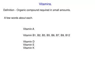
D- VITAMINS. Vitamins are organic compounds required by the body in trace amounts to perform specific cellular functions. They can be classified according to their solubility and their functions in metabolism. Thiamine (vitamin B1)


D- VITAMINS
E N D
Presentation Transcript
Vitamins are organic compounds required by the body in trace amounts to perform specific cellular functions. They can be classified according to their solubility and their functions in metabolism.
Thiamine (vitamin B1) Thiamine pyrophosphate (TPP) is the biologically active form of the vitamin, formed by the transfer of a pyrophosphate group from ATP to thiamine. Thiamine pyrophosphate serves as a coenzyme in the oxidative decarboxylation of α-keto acids.
Distribution of thiamine: Pork, whole grains, and legumes are the richest sources of thiamine. Clinical indications for thiamine: The oxidative decarboxylation of pyruvate and α-ketoglutarate plays a key role in energy metabolism of most cells, but is particularly important in tissues of the nervous system. In thiamine deficiency the activity of these two dehydrogenase reactions is decreased, resulting in a decreased production of ATP, and thus impaired cellular function.
Beri-beri: This is a severe thiamine-deficiency syndrome found in areas where polished rice is the major component of the diet. Signs of infantile beri-beri include tachycardia, vomiting, convulsions, and, if not treated, death. Adult beri-beri is characterized by dry skin, irritability, disorderly thinking, and progressive paralysis.
Wernicke-Korsakoff syndrome: Thiamine deficiency is seen primarily in association with chronic alcoholism and is due to dietary insufficiency or impaired intestinal absorption of the vitamin. Some alcoholics develop the Wernicke-Korsakoff syndrome, a deficiency state characterized by apathy, loss of memory, and a rhythmical to-and-fro motion of the eyeballs.
Riboflavin (vitamin B2) The two biologically active forms are flavin mononucleotide (FMN) and flavin adenine dinucleotide (FAD) formed by the transfer of an AMP moiety from ATP to FMN. FMN and FAD are each capable of reversibly accepting two hydrogen atoms, forming FMNH2 or FADH2. FMN and FAD are bound tightly—sometimes covalently—to flavoenzymes that catalyze the oxidation or reduction of a substrate.
Distribution of riboflavin: Milk, eggs, liver, and green leafy vegetables are good sources of riboflavin. The vitamin is readily destroyed by ultraviolet components of sunlight. Deficiency of riboflavin: Riboflavin deficiency is not associated with a major human disease. Deficiency symptoms include dermatitis, cheilosis (fissuring at the corners of the mouth), and glossitis) thetongue appearing smooth and purplish).
Niacin Niacin, or nicotinic acid, is a substituted pyridine derivative. The biologically active coenzyme forms are nicotinamide adenine dinucleotide (NAD+) and its phosphorylated derivative, nicotinamide adenine dinucleotide phosphate (NADP+).
Distribution of niacin: Niacin is found in unrefined and enriched grains and cereal, milk, and lean meats, especially liver. Niacin can also be obtained from the metabolism of tryptophan. Clinical indications for niacin: Deficiency of niacin causes pellagra, a disease involving the skin, Gl tract, and central nervous system. The symptoms of pellagra progress through the three Ds: Dermatitis, Diarrhea, Dementia, and, if untreated, death.
Treatment of hyperlipidemia. Niacin1 (at doses of 1.5 g/day or 100 times the RDA) strongly inhibits lipolysis in adipose tissue - the primary producer of circulating free fatty acids. Biotin Biotin is a coenzyme in carboxylation reactions, in which it serves as a carrier of activated carbon dioxide.
Distribution of biotin: Biotin is present in almost all foods, particularly liver, milk, and egg yolk. Most American diets contain adequate biotin. Deficiency of biotin: Biotin deficiency does not occur naturaly because the vitamin is widely distributed in food. Also, a large percentage of the biotin requirement in humans is supplied by intestinal bacteria.
Pantothenic acid. Pantothenic acid is a component of coenzyme A, which functions in the transfer of acyl groups. Coenzyme A contains a thiol group that carries acyl compounds as activated thiol esters. Eggs, liver, and yeast are the most important sources of pantothenic arid although the vitamin is widely distributed. Pantothenic acid deficiency is not well characterized in humaris.
Folic acid Folic acid (or folate) plays a key role in one-carbon metabolism, and isessential for the biosynthesis of the purines and the pyrimidline, tnymineiysee. Structure of folic acid The biologically active form of folic acid is tetrahydrofolic acid (THF), which is produced by the two-step reduction of folate by dihydrofolate reductase.
Function of folic acid: Tetrahydrofolate receives one carbon fragments from donors such as serine, glycine, and histidine and transfers them to intermediates in the synthesis of amino acjds, purines and thymidine - the characteristic pyrimidine of DNA. Distribution of folic acid: Folic acid is found in green leafy vegetables, liver, lima beans, and whole grain cereals. Clinical indications: Folic acid deficiency is characterized by growth failure and megaloblastic anemia. The janemia is a result of diminished DNA synthesis in erythropoietic stem cells, a process that requires tetrahydrofolate derivatives.
Folate deficiency: The primary use of the vitamin is in deficiency states that arise from inadequate levels of folate. These can be caused by increased demand (for example, pregnancy and lactation) or by poor absorption due, to pathology of the small intestine, alcoholism, or treatment with drugs that are dihydrofolate reductase inhibitors (for example, methotrexate). A primary result of folic acid deficiency is megaloblastic anemia caused by diminished synthesis of purines and thymidine leading to an inability of cells to make DNA and to divide.
Folate and neural tube defects in the fetus: Early fetal development of the neural tube is critically dependent on the presence of folic acid. Cobalamin (vitamin B12) Vitamin B12 is required in humans for two essential enzymatic reactions: the synthesis of methionine and the isomerization of methylmalonyl CoA that arises from fatty acids with odd numbers of carbon atoms.
When the vitamin is deficient, abnormal fatty acids accumulate and become incorporated into cell membranes, including those of the nervous system. This may account for some of the neurologic manifestations of vitamin B12 deficiency. Structure of cobalamin and its coenzyme forms: Cobalamin contains a corrin ring system that differs from the porphyrins in that two of the pyrrole rings are linked directly rather than through a methene bridge. Cobalt is held in the center of the corrin ring by four coordination bonds from the nitrogens of the pyrrole groups. The remaining coordination bonds of the cobalt are with the nitrogen of 5,6-dimethylbenzimidazole and with cyanide in commercial preparations of the vitamin in the form of cyanocobalamin.
Distribution of cobalamin: Vitamin B12 is synthesized only by microorganisms; it is not present in plants. Animals obtain the vitamin preformed from their natural bacterial flora or by eating foods derived from other animals. Cobalamin is present in appreciable amounts in liver, whole milk, eggs, oysters, fresh shrimp, pork, and chicken. Folate trap hypothesis The effects of cobalamin deficiency are most pronounced in rapidly dividing cells such as the erythropoietic tissue of bone marrow and the mucosal cells of the intestine. Such tissues need the N5-N10-methylene and N10-formyl forms of tetrahydrofolate for the synthesis of nucleotides required for DNA replication. In vitamin B12 deficiency, the N5-methyl form of tetrahydrofolate is not efficiently used.
Clinical indications for vitamin B12: ln contrast to other water-soluble vitamins, significant amounts (4 to 5 mg) of vitamin B12 are stored in the body, As a result, it may take several years for the clinical symptoms of B12 deficiency to develop in individuals who have had a partial or total gastrectomy (who therefore become intrinsic factor-deficient) and can no longer absorb the vitamin.
Pernicious anemia: vitamin B12 deficiency is rarely due to an absence of the vitamin in the diet. It is much more common to find deficiencies in patients who fail to absorb the vitamin from the intestine, resulting in pernicious anemia. The disease is most commonly due to an autoimmune destruction of the gastric parietal cells that are responsibIe for the synthesis of a glycoprotein called intrinsic factor. Normally, vitamin B12 obtained from the diet binds to intrinsic factor in the intestine. The cobalamin-intrinsic factor complex travels through the gut and eventually binds to specific receptors on the surface of mucosal cells of the ileum. The bound cobalamin is transported into the mucosal cell and subsequently into the general circulation, where it is carried by B12-binding protejns.
Lack of intrinsic factor prevents the absorption of vitamin B12, resulting jn pernicious anemia. Pyridoxine (vitamin B6) Vitamin B6 is a collective term for pyridoxine, pyridoxal, and pyridoxamine, all derivatives of pyridine, differing only in the nature of the functional group attached to the ring. Pyridoxine occurs primarily in plants, whereas pyridoxal and pyridoxamine are found in foods obtained from animals. All three compounds can serve as precursors of the biologically active coenzyme, pyridoxal phosphate. Pyridoxal phosphate functions as a coenzyme for a large number of enzymes, particularly those that catalyze reactions involving amino acids.
Distribution of pyridoxine: Good sources of the vitamin are wheat, corn, egg yolk, liver, and muscle meats. The requirement, for pyridoxine is increased by a high intake of protein. Clinical indications for pyridoxine: lsoniazid5 (isonicotinic acid hydrazide), a drug frequently used to treat tuberculosis, can induce a B6 deficiency by forming an inactive derivative with pyridoxal phosphate. Dietary supplementation with B6 is thus an adjunct to isoniazide treatment. Dietary deficiencies in pyridoxine are rare but have been observed in newborn infants fed formulas low in vitamin B6, in women taking oral contraceptives, and in alcoholics. Toxicity of pyridoxine: Neurologic symptoms have been observed at intakes of greater than 2 g/day. Substantial improvement, but not complete recovery, occurs when the vitamin is discontinued.
ASCORBIC ACID (VITAMIN C) Vitamin C has a well documented role as a coenzyme in hydroxylation reactions, for example, hydroxylation of prolyl- and lysyl-residues of collagen. Distribution of ascorbic acid: Citrus fruits, potatoes (particularly their skins) tomatoes, and green vegetables are good sources of vitamin C. CLINICAL INDICATIONS FOR ASCORBIC ACID Deficiency of ascorbic acid: A deficiency of ascorbic acid results in scurvy, a disease characterized by sore, spongy gums, loose teeth, fragile blood vessels, swollen joints, and anemia. Many of the deficiency symptoms can be explained by a deficiency in the hydroxylation of collagen, resulting in defective connective tissue.
Prevention of chronic disease: Vitamin C is one of a group of nutrients, which includes vitamin E and β-carotene, that are known as antioxidants. Supplementation of the diet with these compounds decreases the incidence of chronic disease, such as coronary heart disease and certain cancers. Although vitamins C, E, and β-carotene each show different protective actions, the common chemical property thought to be central to their biologic action is the ability to inactivate toxic oxygen free radicals. Reactive oxygen radicals arise both as a byproduct of normal metabolism and by exposure to sunlight, ozone, tobacco smoke, and other environmental pollutants. Free radicals are known to damage lipid membranes, proteins and cellular DNA; free radicals are thought to play a role in the development of heart and lung disease, cancer, and even aging. Toxicity of ascorbic acid: No acute toxicity has been observed. However, it is known that the oxidized form of ascorbic acid, dehydroascorbic acid, is toxic.
FAT-SOLUBLE VITAMINS These vitamins are released, absorbed, and transported with the fat of the diet. They are not readily excreted in the urine, and significant quantities are stored in the liver and adipose tissue. Vitamin A (retinol) The retinoids are a family of molecules that are related to retinol (vitamin A) and are essential for vision, reproduction, growth, and maintenance of epithelial tissue.
Retinoic acid, derived from oxidation of dietary retinol, mediates most of the actions of the retinoids, except for vision, which depends on retinal, the aldehyde derivative of retinol. Retinoic acid binds with high affinity to specific receptor proteins present in the nucleus of target tissues, such as epithelial cells. Structure of vitamin A: Vitamin A is often used as a collective term for several related biologically active molecules. The term “retinoids” includes both natural and synthetic forms of vitamin A that may or may not show vitamin A activity.
Absorption and transport of vitamin A Transport to liver: Retinol esters present in the diet are hydrolyzed in the intestinal mucosa, releasing retinol and free fatty acids. Release from liver: when needed, retinol is released from the liver and transported to extrahepatic tissues by the plasma retinol-binding protein (RBP). The retinol-RBP complex attaches to specific receptors on the surface of the cells of peripheral tissues, permitting retinol to enter. Many tissues contain a cellular retinol-binding protein that carries retinol to sites in the nucleus where the vitamin acts in a manner analogous to steroid hormones.
Functions of vitamin A Visual cycle: Vitamin A is a component of the visual pigments of rod and cone cells. Rhodopsin, the visual pigment of the rod cells in the retina, consists of 11-cis retinal specifically bound to the protein opsin. Growth: Animals deprived of vitamin A initially lose their appetites, possibly because of keratinization of the taste buds. Bone growth is slow and fails to keep pace with growth of the nervous system, leading to central nervous system damage. Reproduction: Retinol and retinal are essential for normal reproduction, supporting spermatogenesis in the male and preventing fetal resorption in the female. Retinoic acid is inactive in maintaining reproduction and in the visual cycle, but promotes growth and differentiation of epithelial cells; thus, animals given vitamin A only as retinoic acid from birth are blind and sterile.
Maintenance of epithelial cells: Vitamin A is essential for normal differentiation of epithelial tissues and mucus secretion. Distribution of vitamin A: Liver, kidney, cream, butter, and egg yolk are good sources of preformed vitamin A. Yellow and dark green vegetables and fruits are good dietary sources of the carotenes, which serve as precursors of vitamin A. Clinical indications Dietary deficiency: Night blindness is one of the earliest signs of vitamin A deficiency. The visual threshold is increased making it difficult to see in dim light. Prolonged deficiency leads to an irreversible loss in the number of visual cells. Severe vitamin A deficiency leads to xerophthalmia, a pathologic dryness of the conjunctiva and cornea.
Prevention of chronic diseased: Populations consuming diets high in β-carotene show decreased incidence of heart disease and lung and skin cancer. Toxicity of retinoids Excessive intake of vitamin A produces a toxic syndrome called hypervitaminosis A. Amounts exceeding 7.5 mg of retinol per day should be avoided. Early signs of chronic hypervitaminosis A are reflected in the skin, which becomes dry and pruritic, the liver, which becomes enlarged and can become cirrhotic, and in the nervous system, where a rise in intracranial pressure may mimic the symptoms of a brain tumor. Pregnant women particularly should not ingest excessive quantities of vitamin A because of its potential for causing congenital malformations in the developing fetus.
Vitamin D The D vitamins are a group of sterols that have a hormone-like function. The active molecule, 1,25-dihydroxycholecalciferol (1,25 diOH D3), binds to intracellular receptor proteins. The 1,25-diOH D3-receptor complex interacts with DNA in the nucleus of target cells and either selectively stimulates gene expression, or specifically represses gene transcription. The most prominent actions of 1,25-diOH D3 are to regulate the plasma levels of calcium and phosphorus. Distribution of vitamin D Diet: Ergocalciferol (vitamin D2), found in plants, and cholecalciferol (vitamin D3), found in animal tissues, are sources of preformed vitamin D activity.
Ergocalciferol and cholecalciferol differ chemically only in the presence of an additional double bond and methyl group in the plant sterol. Endogenous vitamin precursor: 7-Dehydrocholesterol, an intermediate in cholesterol synthesis, is converted to cholecalciferol in the dermis and epidermis of humans exposed to sunlight. Metabolism of vitamin D Vitamins D2 and D3 are not biologically active, but are converted in vivo to the active form of the D vitamin by two sequential hydroxylation reactions. The first hydroxylation occurs at the 25-position and is catalyzed by a specific hydroxylase in the liver. The product of the reaction, 25-hydroxycholecalciferol or 25-OH D3, is the predominant form of vitamin D in the plasma, and is the major storage form of the vitamin. 25-OH D3 is further hydroxylated at the 1 position by a specific 25-hydroxycholecalciferol 1-hydroxylase found primarily in the kidney, resulting in the formation of 1,25-dihydroxycholecalciferol (1,25-diOH D3).
Function of vitamin D: The overall function of 1,25-diOH D3 is to maintain adequate plasma levels of calcium. It performs this function by (1) increasing uptake of calcium by the intestine, (2) minimizing loss of calcium by the kidney, and (3) stimulating resorption of bone when necessary. Effect of vitamin D on the intestine: 1,25-diOH D3 stimulates the intestinal absorption of calcium and phosphate. 1,25-diOH D3 enters the intestinal cell and binds to a cytosolic receptor. Calcium uptake is enhanced by an increased synthesis of a specific calcium-binding protein. Effect of vitamin D on bone: 1,25-diOH D3 stimulates the mobilization of calcium and phosphate from bone by a process that requires protein synthesis and the presence of PTHT.
Distribution and requirement of vitamin D: Vitamin D occurs naturally in fatty fish, liver, and egg yolk. Clinical indications Nutritional rickets: Vitamin D deficiency causes a net demineralization of bone resulting in rickets in children and osteomalacia in adults. Rickets is characterized by the continued formation of the collagen matrix of bone but incomplete mineralization, resulting in soft, pliable bones. In osteomalacia, demineralization of preexisting bones increases their susceptibility to fracture.
Renal rickets (renal osteodystrophy): This disorder results' from chronic renal failure and thus the decreased ability to form the active form of the vitamin 1,25-diOH cholecalciferol (calcitriol) administration is effective replacement therapy. Hipoparathyroidism: Lack of parathyroid hormone causes hypocalcemia and hyperphosphatemia. These patients may be treated with any form of vitamin D along with parathyroid hormone. Toxicity of vitamin D: Vitamin D is the most toxic of all vitamins. Like all the fat-soluble vitamins, vitamin D can be stored in the body and is only slowly metabolized. High doses (100,000 IU for weeks or months) can cause loss of appetite, nausea, thirst, and stupor.]
Vitamin K Function of vitamin K γ - Carboxyglutamatel: Vitamin K is required in the hepatic synthesis of prothrombin and the blood clotting factors VII, IX, and X. Formation of the clotting factors requires the vitaminj K-dependerit_carboxylation of glutamic acid residues forming a mature clotting factor that contains γ-carboxyglutamate. Interaction of prothrombin with platelets: The Gla residues of prothrombin are good chelators of positively charged calcium ions because of the two adjacent, negatively charged carboxylate groups. The prothrombin-calcium complex is then able to bind to phospholipiqls essential for blood clotting on the surface of platelets. Attachment to the platelet increases the rate at which the proteolytic conversion of prothrombin to thrombin can occur.
Distribution and requirement of vitamin: Vitamin K is found in cabbage, cauliflower, spinach, egg yolk, and liver. There is also extensive synthesis of the vitamin by the bacteria in the gut. Clinical indications Deficiency of vitamin K: A true vitamin K deficiency is unusual because adequate amounts are generally produced by intestinal bacteria or obtained from the diet. If the bacterial population in the gut is decreased, for example by antibiotics, the amount of endogenously formed vitamin is depressed and can lead to hypoprothrombinemia in the marginally malnourished individual. This condition may require supplementation with vitamin K to correct the bleeding tendency. Deficiency of vitamin K in the newborn: Newborns have sterile intestines and cannot initially synthesize vitamin K. Toxicity of vitamin K: Prolonged administration of large doses of vitamin K can produce hemolytic anemia and jaundice in the infant, due to toxic effects on the membrane of red blood cells.
Vitamin E The E vitamins consist of eight naturally occurring tocopherols, of which a-tocopherol is the most active. The primary function of vitamin E is as an antioxidant in prevention of the nonenzymic oxidation of cell components.