Download
1 / 41
420 likes | 483 Vues
Learn about the pathophysiology, risk factors, diagnosis, and management of Compartment Syndrome, as well as mechanisms of joint stability and management of acute joint dislocation. Understand the importance of early intervention and possible complications involved.

E N D
Orthopedic Emergencies:Compartment Syndrome/Acute Joint Dislocation • Ahmad Bin Nasser MBBS, FRCSC • Assistant Professor • Course 451 • KSU
ObjectivesCompartment Syndrome • To explain the pathophysiology of CS • To Identify patients at risk of developing CS • To be able to diagnose and initially manage patients with CS • To be able to describe the possible complications of CS
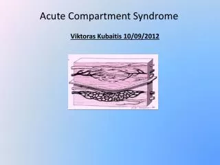
Compartment Syndrome • What is a compartment? • What is the tissue pressure normally? • artery>arteriol>capillary bed (diffusion/exchange)>venule> vein
Compartment Syndrome • Pathophysiology: • risk factor • Elevated tissue pressure • Absence of diffusion at the capillary bed • Cell damage and swelling • Further increase in tissue pressure • Lack of oxygenation • Vicious circle
Compartment Syndrome • Threshold pressure: • 30 mm Hg (rigid) • Less than 30 mm Hg difference between compartment pressure and diastolic pressure (clinically relevant)
Compartment Syndrome • Risk factors (local): • Trauma, crush, fracture (open/closed) • Injection • Bleeding • Prolonged vascular occlusion (reperfusion injury) • Burns • Venomous bite • Intra-osseous fluid replacement • IV fluid extravasation • Tight bandage • Post surgery
Compartment Syndrome • Risk factors (general): • Head injury • Decreased conciseness • Late diagnosis • Hypotension
Compartment Syndrome • Diagnosis: • Early: • Pain!!! • Pain increase with stretching the involved compartment • Presence of risk factor • High index of suspicion • Measurement of compartment pressure is high
Compartment Syndrome • Diagnosis: • Late: • Paresthesia • Paralysis • Pallor • Severely high pressure: • Pulselessness (RARE!)
Compartment Syndrome • Diagnosis: • Tight, woody compartment • Tender compartment • Measurement: • Rarely necessary • Must be done at area of highest expected pressure • May give false low result
Compartment Syndrome • Management: • Initial (undeveloped CS): • Maintain normal blood pressure • Remove any constricting bandage • Keep limb at heart level • Regular close monitoring (15-30 minute intervals) • Avoid nerve blocks, sedation and strong analgesia to obtain patients feed back
Compartment Syndrome • Management: • Fully developed CS • Maintain normal blood pressure • Remove any constricting bandage • Keep limb at heart level • Diuresis to avoid kidney tubular injury if late • Urgent surgical decompression (Fasciotomy)
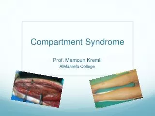
Compartment Syndrome • Fasciotomy: • Releasing the compartment fascia • Allows swollen muscles to expand in volume • Results in decreased compartment pressure • Avoids further damage • Does not reverse already occurred damage • Ideally should be done as soon as diagnosis is made
Compartment Syndrome • Fasciotomy: • Should be done as long as there is still viable tissue • Should not be done if there is no expected viable tissue, Otherwise infection is likely • . Debridement of all necrotic tissue is necessary • Second and third look surgeries are often required • Closure of skin is usually achieved after swelling has subsided • Skin grafting is often required
Compartment Syndrome • Fasciotomy: • Indications: • 6 hours of total ischemia time (ex: arterial embolism) • Significant tissue injury • Worsening initial clinical picture • Delayed presentation with a picture of developed CS • Absolute Compartment pressure >30 mmHg or <30 mm Hg difference from diastolic pressure
Compartment Syndrome • Fasciotomy: • Is a prophylactic procedure • Does not reverse injury to permanently damaged tissue • so better to have a low threshold!
Compartment Syndrome • Complications: • Myonecrosis> myoglobenemia>myoglobinuria> kidney tubular damage • Loss of function of the involved compartment: • Flexion contracture • Paralysis • Loss of sensation
Compartment Syndrome • Complications: • Leg: • Anterior compartment: • Drop foot • Deep posterior compartment: • Clowed toes • Loss of sensation in the sole • Forearm: • Volar compartment: • Volkman contracture
Acute Joint Dislocation • Objectives: • To describe mechanisms of joint stability • To be able diagnose patients with a possible acute joint dislocation • to be able to describe general principles of managing a patient with a dislocated joint • to describe possible complications of joint dislocations in general and in major joints such as the shoulder, hip and knee
Acute Joint Dislocation • Joint stability: • Bony stability • Shape of the joint (ball and socket vs round on flat) • Soft Tissue : • Dynamic stabilizer: Tendons/Muscles • Static stabilizer: Ligaments ± meniscus/labrum • Complex synergy leading to a FUNCTIONAL and STABLE joint
Acute Joint Dislocation • It takes higher energy to dislocate a joint with bony stability than a joint with mainly soft tissue stability • Connective tissue disorders may lead to increased joint instability due to abnormal soft tissue stabilizers. • Dislocation of a major joint should lead to considering other injuries.
Acute Joint Dislocation • At risk group: • Major trauma victims • Athletes and sport enthusiasts • Connective tissue disorder patients
Acute Joint Dislocation • When a joint is subjected to sufficient force in certain directions it might sustain a fracture, a dislocation or a fracture dislocation • Different joints have different force victors that may lead to a dislocation • A joint might dislocate in different directions
Acute Joint Dislocation • A joint dislocation is described by stating the location of the distal segment • Anterior shoulder dislocation: anterior displacement of the humeral head relative to the glenoid • Posterior hip dislocation: posterior displacement of the femoral head relative to the acetabulum
Acute Joint Dislocation • Dislocation: • Total loss of contact between the articular surfaces of the joint • Sublaxation: • partial loss of contact between the articular surfaces of the joint • Acute joint dislocation • Chronic joint dislocation
Acute Joint Dislocation • Diagnosis: • History of a traumatic event ( major trauma or any trauma with the limb in high risk position) • Pain and inability to use the limb • Deformity • Shortening • Malalignment • Malrotation
Acute Joint Dislocation • Diagnosis: • Should check for other injuries (distracting injury) • Should always check the distal neurovascular status. • Should check for compartment syndrome
Acute Joint Dislocation • Diagnosis: • X-rays: • Should be done urgently without delay if dislocation is suspected • Two perpendicular views of the involved joint • Occasionally, special views are required such as the axillary view for shoulder dislocation • X-rays to the joint above and below
Acute Joint Dislocation • Management principles: • Must rule out other injuries • Pain relief • Urgent reduction • Check stability and safety zone • Check neurovascular status after reduction • X-rays after reduction • Protect the joint • Rehabilitation • Follow for late complications
Acute Joint Dislocation • Reduction: • Monitor vitals • IV analgesia (opiod) • IV sedation (to relax the muscles) • Gradual traction to distract the joint • Realignment and rotation to reduce the joint based on direction of dislocation • A palpable clunk well be felt • Check ROM and stability of the joint
Acute Joint Dislocation • Reduction: • Once joint is felt to be reduced, check distal NV status • If it was intact before but not after, farther urgent management is needed • If it was not present before but intact after, check again later to confirm • Observe patients vitals until medications wear out • Stabilize joint and get X-rays
If irreducible or partial reduction only • Urgent closed reduction under general anesthesia and possible open reduction if closed reduction fails • Usually due to • insufficient muscle relaxation • Entrapment of soft tissue
Acute Joint Dislocation • Special considerations: • A fracture dislocation is usually reduced in an open fashion in the operating room • Must confirm concentric reduction on the x-rays, otherwise an open reduction should be performed
Acute Joint Dislocation • Early Complications: • Heterotopic ossification • Neurological injury (reversible or irreversible) • Vascular injury • Compartment syndrome • Osteochondral fracture/injury
Acute Joint Dislocation • Late complications: • Stiffness • Heterotopic ossification • Chronic instability • Avascular necrosis • Osteoarthritis
Acute Joint Dislocation • Special considerations: • Hip joint: • Posterior dislocation is commonest • Major trauma with hip flexed (dashboard injury) • Sciatic nerve injury common • High incidence of late avascular necrosis • An orthopedic emergency!!
Acute Joint Dislocation • Special considerations: • Shoulder dislocation: • common • Anterior dislocation is more common • Patients with seizures prone to posterior dislocation • May cause chronic instability • Can result in axillary nerve injury
Acute Joint Dislocation • Special considerations: • knee dislocation: • Three or more ligaments • Severe (high energy) trauma • May be associated with popletial artery injury---- Limb threatening • Very serious emergency • Needs accurate vascular assessment • May be associate with peroneal nerve injury • May be associated with fracture/ compartment syndrome • Most require surgery either early or late or both
Be safe and alert! Thank you