Download
1 / 29
290 likes | 333 Vues
Eukaryotes are defined by their cellular complexity with organelles like nucleus, mitochondria, and plastids, and a cytoskeleton. Explore the genetic organization and evolutionary theories of eukaryotic cells. Learn about antimitotic alkaloids, cytoskeletal proteins, and protists' diverse characteristics. Discover how Trypanosoma parasites and Chagas' disease pose health risks.

E N D
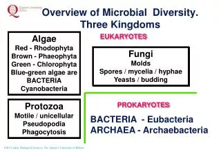
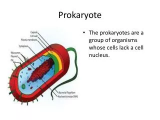
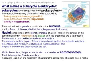
http://phylogeny.arizona.edu/tree/eukaryotes/eukaryotes.html What makes a eukaryote a eukaryote? eukaryotes are distinguished from prokaryotes by the structural complexity of the cells. - characterized by having many functions segregated into semi-autonomous regions (organelles), and by the cytoskeleton. The most evident organelle in most cells is the nucleus, and it is from … this organelle that the eukaryotes get their name. Nuclei contain most of the genetic material of a cell - with other elements of the genome located in mitchondria and plastids (if those organelles are also present). The nucleus is bounded by a membranous envelope. The nuclear envelope is part of the endomembrane system that extends to include the endoplasmic reticulum, dictyosomes (Golgi apparatus) and the plasma membrane that encloses the cell. Within the nucleus, the genes are located on a number of chromosomes. The total amount of DNA in a nucleus measuring less than one hundredth of a millimetre across may stretch to over a metre.
http://phylogeny.arizona.edu/tree/eukaryotes/eukaryotes.html - enzyme dynein - enzyme myosin C&R Fig 7.24 Some antimitotic alkaloids are useful as cancer chemotherapy agents. Taxol{from yew} increases the formation of microtubules and stabilizes them so that there is no free tubulin for the formation of mitotic spindles. Vinblastine{from periwinkel} causes the disassembly of formed microtubules and causes the aggregation of crystaline tubulin. Because tumor cells multiply faster than normal cells they are more susceptible to antimitotic drugs. The cytoskeleton is comprised of a rich array of proteins. The major ones are tubulin (which forms microtubules) and actin (forming microfilaments) and a myriad of interacting proteins which effect movement or create the skeletal architecture of cells.
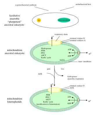
Lynn Margulis The Endosymbiotic Theory of Eukaryote Evolution This idea was promulgated at the turn of the 20th century (Mereschkowsky, 1910) and promoted later by Lynn Margulis (Margulis, 1970).
Right: Spirochetes are masters of movement, corkscrewing about with neither head nor tail. (Photograpgh by Lyn Margulis) Bottom: This contemporary protist, Trichonympha, is pushed through its viscous termite hindgut habitat by thousands of symbiotic spirochete bacteria attaching at the rear. (Photography by David Chase) http://www.earthwise.org/walk/walk_photos/2700.htm
The protistsare a paraphyletic group; those eukaryotes that are notanimals, true fungi or green plants. {this is basically the junk drawer}
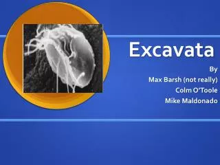
theDiplomonads (two nuclei): theTrichomonads(3 flagella) http://www.nih.gov/news/pr/aug2001/nichd-15.htm Trichomoniasis is caused by infection with … Trichomonas vaginalis. Pregnant women infected with T. vaginalis are more likely to … give birth prematurely to children of low birth weight, … children … are more than twice as likely to be stillborn or to die as newborns… Diplomonadida and Parabasala: lack mitochondria According to what is calledthe "archaezoa hypothesis," these protists are the modern forms of ancient eukaryotic lineages that evolved before the acquisition of the endosymbiotic bacteria that gave rise to mitochondria. Most protistan systematists have recently rejected that hypothesis based on the discovery of mitochondrial genes in the nuclear genomes … the evidence now supports the hypothesis that these protists lost their mitochondria during their evolution from ancestors in which mitochondria were present.
The euglenoids (such as Euglena) are characterized by an anterior pocket from which one or two flagella emerge. Paramylon, a glucose polymer that functions as a storage molecule, is also characteristic of euglenoids. The kinetoplastids have a single large mitochondrion associated with a unique organelle, the kinetoplast, that houses extranuclear DNA. Trypanosoma cause African sleeping sickness, a human disease that is spread by the bite of the tsetse fly Trypanosoma gambiense in a blood smear. http://usa.biologists.com/JCS/gallery.html Immunofluorescence of the trypanosome mutant snl-1 staining the nucleus and the kinetoplast (a large mitochondrion),
Protist: Euglenozoa: Kinetoplasts: Trypanosoma Small animals such as dogs, cats, guinea pigs, rats and opossums can serve as reservoirs for the parasite. T. cruzi generally does not cause the significant disease in animals as it does in people. …There is no known cure for T. cruzi infection in dogs and cats. Because of this and the extreme danger to people, it is generally recommended that infected animals be euthanized. The most effective means of control is the elimination of the kissing bugs. http://www.biosci.ohio-state.edu/~parasite/chagas.html A few species of Trypanosoma are found in the New World. From the standpoint of human health, the most important is Trypanosoma cruzi, causing American trypanosomiasis or Chagas' disease. The vector for Chagas' disease is a "true bug" (Hemiptera) {‘kissing bug’} In the human host, Chagas' disease affects primarily the nervous system and heart. Chronic infections result in various neurological disorders, including dementia, megacolon, and megaesophagus, and damage to the heart muscle. Left untreated, Chagas' disease is often fatal.
the Cilliates - Paramecium - Fig 28.14 macronucleus (approx 50n) controls ‘everyday functions’ & micronuclei - swapped in sexual conjugation - Fig 28.15 the Apicomplexans(sporozoan parasites) Plasmodium - malaria- Fig 28.7 At present, at least 300,000,000 people are affected by malaria globally, and there are between 1,000,000 and 1,500,000 malaria deaths per year. the Dinoflagellates: phytoplankton - Red Tides Pfiesteria piscicida (‘the Cell from Hell!’) Alveolata draws together a group of flagellates (the dinoflagellates), a group of parasites (apicomplexans), and a distinctive group of eukaryotes that move by means of cilia (the ciliates). Alveolates have small membrane-bounded cavities (alveoli) under their cell surfaces. The function of the alveoli is unknown;
Haplotype diversity and linkage disequilibrium at human G6PD: Recent origin of alleles that confer malarial resistance.Tishkoff et al SCIENCE 293 (5529): 455-462 JUL 20 2001Abstract:The frequencies of Low-activity alleles of glucose-6-phosphate dehydrogenase in humans are highly correlated with the prevalence of malaria. These "deficiency" alleles are thought to provide reduced risk from infection by the Plasmodium parasite and are maintained at high frequency despite the hemopathologies that they cause. Haplotype analysis of "A-" and "Med" mutations at this Locus indicates that they have evolved independently and have increased in frequency at a rate that is too rapid to be explained by random genetic drift. Statistical modeling indicates that the A- allele arose within the past 3840 to 11,760 years and the Med allele arose within the past 1600 to 6640 years. These results support the hypothesis that malaria has had a major impact on humans only since the introduction of agriculture within the past 10,000 years and provide a striking example of the signature of selection on the human genome.
PhD - MSU 1986 Alveolata: Dinoflagellates:
What is Pfiesteria ? Pfiesteria piscicida (fee-STEER-ee-uh pis-kuh-SEED-uh) is a toxic dinoflagellate that has been associated with fish lesions and fish kills in coastal waters from Delaware to North Carolina. … dinoflagellates are microscopic, free-swimming, single-celled organisms, usually classified as a type of alga. Although many dinoflagellates are plant-like and obtain energy by photosynthesis, others, including Pfiesteria, are more animal-like and acquire some or all of their energy by eating other organisms. Discovered in 1988 by researchers at North Carolina State University, Pfiesteria piscicidais now known to have a highly complex life-cycle with 24 reported forms, a few of which can produce toxins. … Pfiesteriaonly becomes toxic in the presence of fish, triggered by their secretions or excrement in the water. At that point, Pfiesteria cells shift forms and begin emitting a powerful toxin that stuns the fish … Other toxins are believed to break down fish skin tissue, … As fish are incapacitated, the Pfiesteria cells feed on their tissues … http://www.epa.gov/owow/estuaries/pfiesteria/fact.html
HEADLINE: The 'Cell From Hell' Newsweek August 25, 1997 … what triggers the proliferation of Pfiesteria. In Maryland, environmentalists are blaming the manure runoff from the region's 600 million chickens. In North Carolina, environmentalists say the culprit is mostly runoff from hog farms. … Burkholder's struggles to get North Carolina to recognize Pfiesteria are legion. A new book, "And the Waters Turned to Blood," recounts how corporations and state officials attacked Burkholder's credibility PhD - MSU 1986 HEADLINE:Scary monsters super creepsNew Scientist June 3, 2000 She calls it the career from hell - and she's only half joking. She's been hounded by powerful industries …and watched her funding disappear. North Carolina had tried for years to deny there was a problem, in Maryland action was swift. The governor … asked for medical tests on about 15 people who had been exposed to the toxins. Some of them were diagnosed in the bottom 2 per cent of the US population in their ability to learn and remember. North Carolina was, I think, embarrassed and the state legislature provided generous funding for a new biohazard level 3 lab. With that we are now making good progress …
A ‘target article’ Pfiesteria piscicida and P. shumwayaereportedly secrete potent exotoxins thought to cause fish lesion events, acute fish kills and human disease… Pfiesteria toxins have never been isolated or characterized8. We investigated mechanisms by which P. shumwayae kills fish … Here we show that larval fish bioassays conducted in tissue culture plates fitted with polycarbonate membrane inserts exhibited mortality (100%) only in treatments where fish and dinospores were in physical contact. No mortalities occurred in treatments where the membrane prevented contact between dinospores and fish. … Videomicrography and electron microscopy show dinospores swarming toward and attaching to skin, actively feeding, and rapidly denuding fish of epidermis. We show here that our cultures of actively fish-killing P. shumwayae do not secrete potent exotoxins; rather, fish mortality results from micropredatory feeding. …
A digression on your News & Views assignment A cute, catchy title Establishing a problem Certain episodes of mass fish mortality in coastal waters off the eastern United States have been ascribed to a planktonic organism called Pfiesteria. There are now fresh clues to how these fish are killed. In James Powlik's novel Sea Change1, a colony of mutant cells called Pfiesteria grows into a carpet-like monster stretching over several square miles of the Strait of Juan de Fuca, a body of water separating the west coast of the United States and Canada. Its slow movement south threatens the residents of Seattle with a nightmarish fate — death following inhalation or ingestion of a toxin produced by the microbes. The good news is that the Sea Change story is fiction; the bad news is that Pfiesteria is fact. But what is the truth about this 'phantom of the ocean'?It is thought that Pfiesteria, which belongs to a group of planktonic organisms called dinoflagellates, releases a toxin that ultimately kills fish2.But using simple methods, Vogelbein and his colleagues (page 967 of this issue3) conclude that the toxin is not released into the environment and that the organism kills by direct methods. continued … Luring in the reader with a question (below) & a teaser (above) The ‘established wisdom’’ The ‘bottom line’’ on how the target article is going to change the ‘established wisdom’’
In reality, the presence of Pfiesteria has not been documented along the US west coast but rather along the eastern seaboard, with a range from Delaware to Alabama4. It has been associated with massive fish kills, especially off North Carolina2. Because of the harm that results when Pfiesteria multiplies to high numbers, it is categorized with other plankton known to cause harmful 'blooms'. Many of these bloom-forming organisms produce potent toxins, so the assumption that Pfiesteria releases a toxin is not unfounded. Moreover, the population explosions of various single-celled plankton, including diatoms and dinoflagellates, are frequently associated with the production of nerve toxins — saxitoxin, brevetoxin, maitotoxin and the like5 — that are damaging when ingested by higher organisms. These toxins are the key for researchers wishing to unlock the secrets of harmful algal blooms. By knowing the chemical identity of the toxins, highly specific detection methods can be developed to track the location of blooms and to discover where they originate, how they spread and when they are most toxic. The impact of harmful algae on living creatures, such as seabirds, marine mammals and even humans, can be assessed by determining the levels of toxin in their tissues and body fluids. But first the toxins must be isolated from the clusters of cells that release them into the environment. This is typically done by picking a single suspect cell from the mixture of microbes in a water sample from an estuary or ocean during a harmful event, and allowing it to multiply in the laboratory. Chemical constituents are then extracted from several litres of the cells, and toxic components are identified using a bioassay, a test that assesses whether a particular fraction is poisonous, usually to a fish or mouse. For almost a decade, researchers have tried unsuccessfully to identify, from many litres of isolated Pfiesteria cells, a toxin that could explain the mass mortalities observed in the wild6. continued … The basic story before the target article
Vogelbein et al.3 and — in a related study, with some authors in common — Berry et al.7 have taken a different approach to the quest for a toxin released by Pfiesteria. First, they grew Pfiesteria in the lab and determined that the cultures could kill fish through direct contact. Then they separated the cells from the rest of the material using several methods, including filtration, centrifugation and dialysis (Fig. 1). They theorized that, if Pfiesteria cells release a toxin into sea water, this should be present in the cell-free fractions. But the experiments showed that the cell-free fractions do not cause fish death, and that Pfiesteria is lethal only when cells are in direct contact with the fish. The researchers therefore concluded that a toxin is not released into the surrounding sea water. Figure 1 The dialysis technique used by Vogelbein et al.3 If they are not killed by a toxin, how do fish exposed to Pfiesteria die? It has been known for many years that Pfiesteria cells extend a suction-cup-like appendage called a peduncle to digest fish tissue2. Vogelbein et al. go a step further and propose that fish die because Pfiesteria literally sucks the life out of them. It attaches to fish skin using the peduncle, extending finger-like protrusions called filopodia, then ingests cell matter from the fish. This parasitic feeding behaviour by Pfiesteria is detailed in high-magnification microscope images in Vogelbein et al.3, and in a video clip available in their Supplementary Information. continued … A nice, simple summary of what the authors did & found
The new work promises to bring us closer to unveiling the true nature of this phantom of the ocean. Many loose ends remain, however, and it is possible nonetheless that a toxin is involved. Humans have suffered from memory impairment thought to stem from exposure to Pfiesteria8. Are these memory problems caused by a toxin, in aerosol form, produced by another organism that often coexists with Pfiesteria, as Berry et al.7 suggest? Do the rod-shaped granules that Vogelbein and colleagues identified in Pfiesteria (see Fig. 4 on page 969) contain toxins that are released only after the peduncle becomes attached? Do only certain Pfiesteria isolates produce toxins, as Burkholder et al.9 have proposed, indicating that the Pfiesteria studied by Vogelbein, Berry and their colleagues were simply non-toxic strains? Can contamination by a fungus or other microbe explain why Pfiesteria cultures routinely grown in the presence of fish do indeed appear to produce a toxin10? Assembling these pieces of the complex puzzle posed by Pfiesteria will require exceptional cooperation among researchers of differing expertise. The opportunity to discuss the outstanding questions and to establish new collaborations will engage scientists at the Tenth International Conference on Harmful Algal Blooms, to be held in St Petersburg, Florida, this October. References 3.Vogelbein, W. K. et al. Nature418, 967-970 (2002); What questions are left unanswered & what needs to be done next? You only need to cite the target article
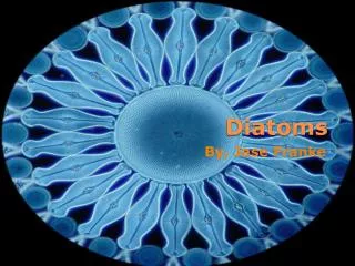
the Water Molds (not fungi because cell walls cellulose, not chitin) ‘ick’ on fish - Fig 28.16 downy mildews on plants: potato blight, vine root rot the Diatoms: - Fig 28.17 have unique glasslike walls consisting of hydrated silica embedded in an organic matrix. Each wall is in two parts that overlap like a shoe box and lid – most of the year, diatoms reproduce asexually by mitotic cell divisions, with each daughter cell receiving half of the cell wall of its parent and regenerating a new second half. Most golden algae are unicellular, their cells biflagellated. Some species are mixotrophic, absorbing dissolved organic compounds or ingesting food particles and bacteria by phagocytosis, Many of the ‘seaweeds’ are brown algae (ex: kelp). Stramenopila includes several heterotrophic groups as well as a variety of photosynthetic protists (algae). The term stramenopila refers to the numerous fine, hairlike projections on the flagella that are characteristic of these organisms
Unlike other eukaryotic algae, red algae have no flagellated stages in their life cycle. They are reddish because of an accessory pigment called phycoerythrin also found in cyanobacteria.
Fig 28.24 Molecular systematics and cellular morphology leave little doubt that green algae and land plants are closely related. Some systematists advocate an expanded kingdom, the Viridiplantae.
Amoeboid things ‘A diversity of protists that use pseudopodia for movement and feeding Little is known about their phylogeny, although it is clear that they represent several distinct eukaryotic lineages. Because of taxonomic uncertainties, we have not included these protists on the phylogenetic tree the Rhizopods (amoebas) ‘a polyphyletic group’ The cytoskeleton of microtubules and microfilaments functions in amoeboid movement. Entamoeba histolytica causes amoebic dysentery. Actinopod means "ray foot," a reference to the slender pseudopodia that radiate from these protists. Each axopodium is reinforced by a bundle of microtubules covered by a thin layer of cytoplasm. Most actinopods are planktonic, and their projections place an extensive area of cellular surface in contact with the surrounding water, Foraminiferans, or forams, are named for their porous shells. Almost all are marine, most species live in the sand or on rocks and algae, but some are also abundant in plankton, deriving nourishment from the photosynthesis of symbiotic algae that live within the shells. ?
Acellular Plasmodialslime molds - diploid - Fig 28.29 http://www.ucmp.berkeley.edu/protista/slimemolds.html Plasmodial slime molds, like Physarum shown here, are formed when individual flagellated cells swarm together and fuse into one large bag of cytoplasm with many diploid nuclei. Dictyostelida (Cellular Slime Molds) – haploid Fig 28.30 Although the feeding stage of the life cycle consists of solitary cells that function individually, when food is depleted the cells form an aggregate that functions as a unit. Poses questions about what it means to be an individual organism. Serves as a model for evolution of multicellularity & cooperation Mycetozoa translates as "fungus animals," though these protists are neither fungi nor animals.
C&R Fig 28.30 http://www.ucmp.berkeley.edu/protista/slimemolds.html the cellular slime molds, spend most of their lives as separate single-celled amoeboid protists, but upon the release of a chemical signal {cAMP}, the individual cells aggregate … they provide a comparatively simple and easily manipulated system for understanding how cells interact to generate a multicellular organism.
Ennis et al 2000. PNAS 97:3292-3297. One of the problems … is that in the wild, parasiticforms of Dictyostelium should arise that form only spore cells. Such parasites, not having to tithe20% to the stalk cells, would increase in the population and eventuallyimpede the cooperativeness that leads to fruiting body formation. Dictyostelium amoebae feed on bacteria during growth, but when the food supply is exhausted, they aggregate to form a … cylindrical slug … about 80% of the amoebae form spore cells,whereas 20% form stalk cells. Spore cells germinate to give riseto the next generation,but stalk cells die. The stalk cells havebeen described as altruistic, sacrificing themselvesfor the improveddispersal of spores.
Altruism and social cheating in the social amoeba Dictyostelium discoideumStrassmann JE, Zhu Y, Queller DCNATURE 408 (6815): 965-967 DEC 21 2000Abstract:The social amoeba, Dictyostelium discoideum, is widely used as a simple model organism for multicellular development(1,2), but its multicellular fruiting stage is really a society. Most of the time, D. discoideum lives as haploid, free-living, amoeboid cells that divide asexually. When starved, 10(4)-10(5) of these cells aggregate into a slug. The anterior 20% of the slug altruistically differentiates into a non-viable stalk, supporting the remaining cells, most of which become viable spores(3-5). If aggregating cells come from multiple clones, there should be selection for clones to exploit other clones by contributing less than their proportional share to the sterile stalk. Here we use microsatellite markers to show that different clones collected from a field population readily mix to form chimaeras. Half of the chimaeric mixtures show a clear cheater and victim. Thus, unlike the clonal and highly cooperative development of most multicellular organisms, the development of D. discoideum is partly competitive, with conflicts of interests among cells. These conflicts complicate the use of D. discoideum as a model for some aspects of development, but they make it highly attractive as a model system for social evolution. Continued …
In social situations, opportunities arise for some individuals to take advantage of others. This happens in wild populations of the social amoeba Dictyostelium discoideum. Most of the organisms studied by developmental biologists — the fruitfly Drosophila, for instance — arise from a fertilized egg. So, apart from the mature reproductive cells, the cells from which these organisms are made are genetically identical. No cell has a genetic advantage over the others, and any variants that do arise are excluded from the germ line. Because of the lack of genetic diversity among their cells and the presence of a germ line, cheating by individual cells is restricted in most multicellular organisms. But there is another route to multicellularity, taken by organisms such as Dictyostelium and Myxococcus, a group of social bacteria. As they describe on page 965 of this issue1, Strassmann and colleagues have studied the curious cooperative behaviour of Dictyostelium, and find that some genetic variants cheat on their partners during the process of spore formation (Fig. 1). continued …
replicating DNA molecule Any modern organism is a Russian doll ofactually or potentially reproducing units. Selection can, in principle,act on each such unit. We will, of course, not see its operationin most modern contexts, because conflict between units of selectionare evident only when a chimera isformed. Chimeras that pit one unit of selection against another arise in specific ecological contexts, as exemplified by … slimemolds … Vol. 96, Issue 16, 8801-8803, August 3, 1999CommentarySlime molds, ascidians, and the utility of evolutionary theory Leo W. Buss {on Ennis et al 2000. PNAS 97:3292-3297.} Consider a social insect colony. The colony as a unit reproduces. The ants within a colony reproduce. The cells within eachant reproduce. The mitochondria with each cell reproduce, as does the chromosome of the mitochondria and those of the nucleus. Andwithin those chromosomes are likely transposable elements … They also can arise by mutationduring the life of an individual, as any oncologist knows well.