Lymphatic/Hematopoetic System IPM 2
Lymphatic/Hematopoetic System IPM 2. Scott E. Smith M.D., Ph.D. 12-10-02. Objectives. Student should be able to … describe location, size, consistency, and other attributes of lymphadenopathy identify common clinical scenarios involving lymphadenopathy

Lymphatic/Hematopoetic System IPM 2
E N D
Presentation Transcript
Lymphatic/Hematopoetic SystemIPM 2 Scott E. Smith M.D., Ph.D. 12-10-02
Objectives Student should be able to … • describe location, size, consistency, and other attributes of lymphadenopathy • identify common clinical scenarios involving lymphadenopathy • identify the signs and symptoms of anemia • define the signs and symptoms of bleeding and coagulation disorders
Overview • This is a short lecture! • A major goal is to synthesize the lymphatic system as a whole…lymph node regions have been discussed individually by specific site…i.e., head, neck, and abdomen, but not put together for systemic illness such as lymphoma. • We will also discuss the signs and symptoms of anemias, leukemias, bleeding disorders, and coagulation disorders
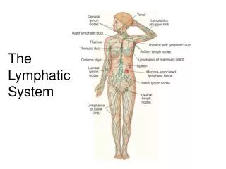
Lymph Node Examination • Head/neck • Axillary • Inguinal/femoral
Head and Neck Nodes • Preauricular • Posterior auricular • Occipital • Tonsillar • Submandibular • Submental • Superficial cervical • Posterior cervical • Deep cervical • Supraclavicular
Axillary • A pectoral (anterior) • L lateral • P posterior • C central • Ap apical
Inguinal/ Femoral • Horizontal group • Vertical group
Descriptors of Lymphadenopathy • Location…obvious • Mobility • Size • Texture • Shape • Tender/non-tender • Associated erythema or warmth…signs of inflammation
Spleen • Left upper quadrant • Palpation most specific for detecting enlarged spleen (89-99% specificity) • Spleen palpable to umbilicus is suggestive of hematologic pathology • Percussion is non-sensitive (dullness in Traube’s space) but can be specific in non-obese patients
Case • 28 yo man presents with c/o fevers, night sweats and 30 pound weight loss. He develops pruritis when he showers. He also has noted some enlarged “glands” in his neck and armpits. On lymphatic exam he has the following:
Case • painless lymphadenopathy in anterior axilla and anterior cervical as well as supraclavicular areas bilaterally. • Lymph nodes are not tender, freely mobile and no associated inflammation. They are ovoid (grape-shaped) and measure 2 x 3 cm. There is no splenomegaly by palpation or percussion.
Differential Diagnosis • Lymphoma • Infection • Cancer—metastatic • Granulomatous disease
Dyspnea on exertion Palpitations Angina pectoris Intermittent claudication Headache Syncope anorexia Dizziness/vertigo Nausea Cold intolerance Amenorrhea Decrease libido/impotence Anemia- Signs/Symptoms
Anemia • Blood loss • Hemolysis/sequestration • Deficiencies • Decreased production
Symptoms • Symptoms based on acuity of HgB drop • Acute blood loss usually creates rapid onset of symptoms • Slow drop in HgB may lead to fewer symptoms
Anemia of Acute Blood Loss • Trauma or GI tract loss most common • Menstrual/vaginal loss • Urinary tract • Nosebleeds leading to anemia, but not because of it! • Tachycardia and hypotension are common findings • History helps the most for these
Hemolysis and Sequestration • Causes for hemolytic anemias include: • Autoimmune • Drug induced • Cell membrane disorders • Hereditary • Splenomegaly can lead to sequestration of blood cells
Scleral Icterus • Yellow sclera • Can be seen in hemolysis
Deficiencies • Iron deficiency anemia is most common worldwide and in US-spoon nails and pica • Megaloblastic anemias caused by B12 or folate deficiencies-paresthesias and diarrhea • Smooth tongue/glossitis
Signs and Symptoms of Coagulation Disorders • Bleeding • Ecchymoses • Petechiae • Hemarthroses • Hematomas
Platelets versus Coags • Petechiae—platelets low or dysfunctional • Ecchymoses, hematomas, hemarthroses—seen more frequently with low clotting factors or dysfunction • Bleeding can be seen with either