Download

1 / 11
110 likes | 671 Vues
Diagnostic Accuracy of Rubidium-82 Myocardial Perfusion Imaging using PET CT. Normal Rubidium-82 Myocardial Perfusion PET-CT Study. Abnormal Rubidium-82 Myocardial Perfusion PET-CT Study. Limitations. Limitations.

E N D
Diagnostic Accuracy of Rubidium-82 Myocardial Perfusion Imaging using PET CT
Limitations • May underestimate the anatomical extent of CAD in some patients with multivesseldisease. • Subclinical vessel disease may have normal perfusion studies. • Not all patients with normal perfusion study get CTA or Cath. Adds bias to the true Negative group
Advantage of ECG-gated PET • Assess LV function at rest and during peak stress • Healthy pts: LVEF increases during peak vasodilator stress • Patients with left main or 3 vessel CAD show decrease in LVEF at peak stress even in the absence of perfusion abnormalities
Sensitivity • 93% in single vessel disease. • 92% in multivessel disease. • May have a slight bias as many normals don’t have correlative coronary angiography. • MDCT angiography shows high degree of correlation with Rubidium-82 Myocardial Perfusion PET-CT.
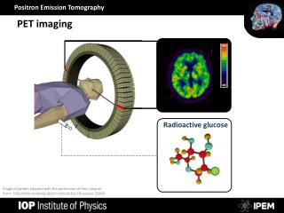
Integrated PET/CTA study: • Noncalcified plaque (arrow) in proximal LAD with 50%–70% stenosis. However, rest and peak stress myocardial perfusion PET study (bottom left panel) demonstrates only minimal inferoapical ischemia.
Conclusion • Myocardial perfusion PET-CT: • high sensitivity for detection of CAD. • applicable to both men and women, nonobese and obese patients. • Patients with single-vessel as well as multivessel coronary disease. • Rest and stress imaging is completed in approximately 25 min.