Download
1 / 93
930 likes | 1.17k Vues
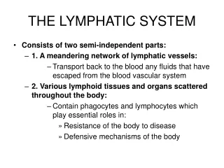
The Lymphatic System and the Blood. Overview. Why needed? Origin: Blood vessels form from mesoderm Blood produced 2 wks after vessels are formed, during the 5 th week of life. What is blood?. Connective tissue? Different from others Matrix not a solid or semi-solid material

E N D
Overview • Why needed? • Origin: • Blood vessels form from mesoderm • Bloodproduced 2 wks after vessels are formed, during the 5th week of life
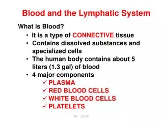
What is blood? Connective tissue? Different from others • Matrixnot a solid or semi-solid material • Matrix of blood is plasma • watery substance • Yellowish • 90% Water • 7% protein • 1% minerals • 2% other materials incl. atmospheric gases, chem signals, and nutrients
More on plasma Contains: • Atmospheric gases: • oxygen, carbon dioxide, and nitrogen • Comprises 55% of blood volume
Formed elements (= Cellular components) Remaining 45% of blood volume: • Erythrocytes (RBCs) • Leukocytes (WBCs) • Thrombocytes (platelets)
Hematocrit Calculates the volume of red blood cells making up the blood Included in a CBC FYI: CBC (on medical shows) = complete blood count
Complete Blood Count includes… • Hematocrit • The number of RBCs • The number of WBCs • The total amount of hemoglobin in the blood Also provides information about the following measurements: • Average red blood cell size (MCV) • Hemoglobin amount per red blood cell (MCH) • The amount of hemoglobin relative to the size of the cell (hemoglobin concentration) per red blood cell (MCHC) • The platelet count is also usually included in the CBC.
Can you answer these questions? • What is the blood composed of? • Why is the blood unlike any other connective tissue? • What does a hematocrit tell you?
Red Blood Cells • No mature nucleus (lost in dev.) • No DNA, so…. • Use enzymes to carry out their tasks • Reticulocytes (immature RBC) – have mesh-like network of rRNA… become mature in ~24 hours • Live max 120 days • No way to repair & replace damaged cellular components • Appear red b/c of hemoglobin • Contains iron facilitates transport of O2 and CO2 • 4.8 million RBC/mm3 in women • 5.4 million RBC/mm3 in men
Blood Type • Genetic • Determined by the antigens on the surface of the RBC membrane • A,B,O blood group system most common (30 possible in full blood type classification!) • Blood will attack “non-self” • Important to match blood types for transfusions • Therefore… • AB universal acceptor • O universal donor-has no proteins on the membrane
Rh Factor • The “D” protein • Most are positive (depends on geography) • If a woman is negative and conceives with a positive man, problems can arise— erythroblastosisfetalis
This can lead to anemia, a condition marked by weakness and fatigue. Severe anemia can lead to heart failure and death. The breakdown of RBC leads to the buildup of bilirubin which can lead to jaundice and brain damage.
Prevention of erythroblastosisfetalis • Treat negative mothers with Rhogam, a preventative measure • Prevents formation of antibodies to Rh molecule • Given whenever there is a possibility of fetal blood mixing with maternal blood following childbirth, abortion, miscarriage, prenatal testing. • Once sensitized the woman will always react against Rh+ cells
Can you answer these questions? • How are RBCs different from most other cells? • How does the lack of a nucleus affect RBCs lifespan? • What is hemoglobin and what does it do? • Why are RBCs red? • What is blood type? What do the different blood types mean? • Why is it dangerous for an Rh- woman to have an Rh+ baby?
White Blood Cells • WBC : RBC ratio = 1 : 500 or 1000 • Use blood, lymph to move from bone marrow to the tissues • 5 types (differential WBC count measures them) • Neutrophils (Most abundant) • Lymphocytes • Eosinophils • Monocytes • Basophils (Least abundant)
WBC’s • Granulocytes Agranulocytes AKA : Mononuclear Noticeable granules that produce specialized secretions for fighting infection Lack visible granules in cytoplasm Nucleus is polymorphic, lobed, unusually shaped Monocytes Lymphocytes Eosinophils, basophils, neutrophils T & B cells
Neutrophils • Granulocyte • Most common WBC • Nucleus = 2-5 lobes • Found in the blood • First responders in the inflammation response due to environmental exposure, some cancers, bacterial infection • Predominant cell in pus
Eosinophils • Granulocyte • 5% of WBCs • Bi-lobed nucleus • Combats parasitic infections (protists, worms) • Secretions produced related to allergies • Normally in thymus GI, ovaries, testes, spleen, uterus, lymph nodes • NOT in lungs, esophagus, or skin if found here, indicates disease/pathology
Basophils • Granulocyte (least common) • Susceptible to basic dyes • Large, bi-lobed nucleus (similar to mast cells) • Granules obscure the nucleus • “Bas-ically all granules” • Involved in allergies. • Stores, secrete histamine & heparin (anticoagulant) • Found where allergic reactions are taking place
Agranulocytes • Lymphocytes • “Immune” cells: • NK (natural killer) cells (no prior activation needed) • T lymphocytes (mature in thymus) • Helper: direct immune response • Cytotoxic: release cytotoxin to kill pathogen infected cells • B lymphocytes (mature in bone marrow): Use antibodies to neutralize pathogens
Monocytes • Agranulocyte • Largest of WBCs • - shaped nucleus • Mono = kissing = Love = heart • Many vesicles in cytoplasm for processing pathogens • Perform phagocytosis- uptake & digestion of pathogens • Fragments of “eaten” pathogen signal T-lymphocytes to the area
Platelets Cell fragments derived from larger cells called megakaryocytes. • Have “sticky” proteins • Reduce blood flow to an affected area. • Reduce blood loss • Sensitive to many types of hazardous chemicals and pollutants
Can you answer these questions? • Describe the characteristics & functions of all granulocytes, agranulocytes, and platelets. • Compare and contrast the structure & function of RBCs and WBCs • Why are platelets called the “Band-Aids” of the blood?
Red Blood Cell Function • Carry oxygen from the lungs to the body • Carry carbon dioxide from the body to the lungs
Alveoli-where gas exchange happens in the lungs • RBC in the capillaries that surround the alveoli oxygen enters • Only if the partial pressure of oxygen outside is higher than inside • In cytoplasm of RBC oxygen binds to Hemoglobin
Hemoglobin • Four oxygen molecules bind to hemoglobin (w/ the iron) • Carries CO2 also; • binds to a different area than O2 • Percent saturation: • amount of oxygen that is dissolved in a solution of hemoglobin molecules • O2sats = 98% or above • Similar to with myoglobin in muscle • Greater affinity for oxygen • Hemoglobin collects oxygen a low partial pressures
In the tissues the oxygen is released and carbon dioxide enters the RBC, binds to Hemoglobin. • Partial pressures of the gases must appropriate • Some cellular wastes stimulate the release of the oxygen from the hemoglobin • Allows RBC to give more O2 to tissues w/ high metabolic needs
Carbon Dioxide Carried 3 ways in the blood 1. Carried in the blood as a gas (10%) 2. Binds to empty hemoglobin: carbaminohemoglobin 3. As a bicarbonate ion (HCO3-) • CO2 can dissolve in water, forming bicarbonate ion • Dissolves in the blood plasma • Carbonic anhydrase: enzyme in RBC that stim’s the formation of carbonic anhydrase, which dissociates to form bicarbonate ions and H+ ions • Eventually excreted
Movement of gases • Diffusion: High concentration low concentration • For Oxygen: • Partial pressure is higher in blood than in tissues • For Carbon Dioxide: • Partial pressure is higher in tissues than in blood
Carbon dioxide intoxication • Occurs when the CO2 is extremely high in the environment or the blood • Acute: high levels in the air • Subacute: toxicity caused by the body’s failure to eliminate carbon dioxide • Decreases blood’s pH (what kind of acid does CO2 form when it dissolves in water?) • Carbonic acid!
Can you answer these questions? • What is the purpose of RBCs? • Where does oxygen bind to the Hb molecule? • Where does Hb collect oxygen? Then what happens? • Describe the partial pressures that must be present for oxygen to diffuse from RBC to tissues and for carbon dioxide to move to the cells?
White Blood Cell Function • In general: • Fight infections & disease • Granulocytes: • granules of toxic chemicals that kill microorganisms • regulate reactions to foreign materials in the body
Neutrophil function • Pass through capillaries to tissues to with infections. • Attracted to affected areas by factors secreted by damaged cells/tissues • Stick to injured tissues, use phagocytosis to engulf remains of bacteria and damaged cells • Secretes antibiotics-harms/kills bacteria • Secretes other chemicals that stim. • Inflammation ↑ blood flow to the area & ↑ WBC concentration
Eosinophil function • Secretions defend against parasitic infections esp. protists & worms • ↑ in eosinophils = parasitic infection • Granules contain major basic protein to kill the parasites • Secrete chemicals associated w/ allergies
Basophil function • Secrete histamine stim the immune response • Overproduction of histamine runny nose, sneezing, watery eyes • Mast cells (special kind of basophil) • Cause inflammation of tissues • Secrete chemical that attract neutrophils • Found in walls of small bl. vessels
Monocyte function • Clear granules give cytoplasm a grey appearance • When they leave the bone marrow they become either: • Circulating monocytes • Detect infections in blood • Bone growth & maintenance • Tissue monocytes (macrophages) • Remove dead cells • Attack microorganisms that are difficult to kill (fungi)
Lymphocyte function • Stay tuned! We’ll talk about it later….for now, they carry out most of the duties of the immune system
Can you answer these questions? • Which WBC is in charge of engulfing bacteria? • Which WBC is in charge of protecting us from parasites? • Which WBC differentiates into cells that assist in bone growth and maintenance or are macrophages that protect against fungal infections? • Which WBC secretes major basic protein?
Platelets’ function • Blood clotting • Platelets adhere to injured area • Activation of blood clot formation • Important that clot forms by injury only • Intact cells secrete prostacyclin(prevents platelet activation)
Clotting Cascade (simplified) 1.) BV damaged, releases “distress chemicals” 2.) Clotting factors stim. other factors that indicates presence of damaged tissues a.) plateletsstick to damaged tissues & each other b.) Platelets secrete prothrombin activator & Ca2+ • Catalyze conversion of prothrombinto thrombin c.) Thrombin causes fibrinogen fibrin d.) Fibrin forms a sticky mesh that adheres to thrombocytes and other blood components (clot) • Clot forms a barrier that prevents blood loss & impedes the passage of microorganisms into tissues • Calcium ions = catalyze PT to T • Vitamin K = synthesis of clotting factors Prothrombin Fibrinogen Fibrin Thrombin
Why is the cascade so complicated? • So the blood doesn’t clot unintentionally! • They aren’t permanent • Plasminogenplasmin (digests fibrin and dissolves a clot) • Healthy cells near the clot secrete TPA (tissue plasminogen activator)dissolves fibrin as well.
Can you answer these questions? 1.) What is the purpose of prostacyclin? 2.) What is the purpose of a clot? 3.) What are the steps of the clotting cascade? 4.) What is the role of calcium and vitamin K in clot formation? 5.) Why is the clot cascade so complex? 6.) What do plasmin and tissue plasminogen have in common? What’s the difference?