Download
1 / 12
120 likes | 325 Vues
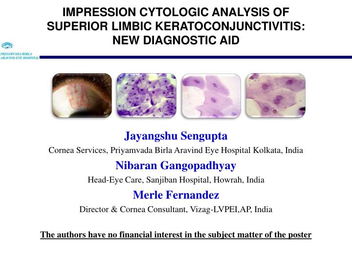
Jayangshu Sengupta Cornea Services, Priyamvada Birla Aravind Eye Hospital Kolkata, India Nibaran Gangopadhyay Head-Eye Care, Sanjiban Hospital, Howrah, India Merle Fernandez Director & Cornea Consultant, Vizag-LVPEI,AP, India

E N D
Jayangshu Sengupta Cornea Services, Priyamvada Birla Aravind Eye Hospital Kolkata, India Nibaran Gangopadhyay Head-Eye Care, Sanjiban Hospital, Howrah, India Merle Fernandez Director & Cornea Consultant, Vizag-LVPEI,AP, India The authors have no financial interest in the subject matter of the poster
Conjunctival impression cytology • Simple, noninvasive technique • Sample the superficial layers of epithelium for analysis. • Gold standard procedure for analysis of squamous metaplasia and goblet cell loss. • Light microscopy- remains most commonly used in clinical practice. • Superior Limbic Keratoconjunctivitis • Disorder of unknown etiology • Role of impression cytology as a diagnostic modality remains largely unknown.
To demonstrate the morphological changes induced in the conjunctival epithelium in presence of SLK using the process of impression cytology. Prospective, non-comparative, observational case series 50 eyes(26 patients) were analyzed 2 eyes with inadequate samples were not analyzed January 2008 – December 2008
Diagnostic criteria Localized superior bulbar congestion with conjunctivochalasis +/- filament Localized punctuate Rose Bengal staining Papillary changes over upper tarsal conjunctiva Bilateral involvement. Exclusion criteria Use of contact lens Schirmers II value<5mm Prior steroid therapy-topical/systemic (within previous 3 months) Evaluation Detailed history Slit lamp examination Schirmer test Tear film break up time Rose Bengal staining Fluorescein stain score Systemic examination
Standard technique of impression cytology • Haematoxylin- PAS staining • Grossly labeled as normal/abnormal • epithelial cell morphology • cohesion of cells • keratinization • nuclear characteristics • goblet cell density. • Abnormal specimens graded • Tsengs classification system (Scheffer C.G.Tseng Ophthalmology 92:728-733,1985) Goblet cells Normal epithelial pattern Abnormal Impression
Age Range was found to be 36 to 55 yrs • M:F ratio of 1:12 • Co morbidity factors were found in 4 patients Hypothyroid Rheumatoid Arthritis 1 patient each Post Lasik High Prolactin level • All samples from superior bulbar portion of conjunctiva demonstrated Grade V squamous metaplasia
Increase in cell size • Lack of cohesiveness with increased intercellular spaces and folded edges • Keratinisation • Complete absence of goblet cells • Pyknotic nucleus
Snake chromatin • 100% • Due to alteration in the nuclear and cytoplasmic skeleton Snake like chromatin Knop E, Reale E Fine structure and significance of snake like chromatin in conjunctival epithelial cells. Invest. Ophthalmol. Visual Sci 35:711, 1994 Margarita Calonge, Yolanda Diebold, Victoria Saez et al Impression cytology of the ocular surface: a review Experimental Eye Research78:457, 2004
Multinucleation (100 perent) Balloon degeneration (96%) Multinucleation Balloon degeneration
Multilobulated nuclei (70%), Spindle configuration (68%) Dumb-bell configuration (60%). Multilobulation Spindle Configuration Dumb- bell Dumb- bell
Hallmark of squamous metaplasia from histopathological specimens • Extensive nuclear pyknosis • Condensation of masses of chromatin • The shrinkage of the nuclear envelope from its surrounding cytoplasm • Though electron microscopy of histological samples in patients with SLK better demonstrates such nuclear changes, conjunctival impression cytology in SLK conclusively demonstrates • Localized squamous metaplasia of high grade • Complete lack of goblet cells • Characteristic nuclear changes like balloon degeneration, condensed chromatin, snake like chromatin, multi lobulated and strangulated nuclei • Formation of snake like chromatin is characteristic • Probably an indicator of mechanical stress on the ocular surface like blink related micro-trauma. Barry Collin, Peter C. Donshik, S. Arthur Boruchoff, et al. The fine structure of nuclear changes in superior limbic keratoconjunctivitis. Invest. Ophthalmol. Visual Sci 17(1):79, 1978
To conclude, impression cytology is a simple, noninvasive method of demonstrating the underlying pathological changes and thereby becomes an important tool in diagnosing SLK.