
RADIO ISOTOPES
E N D
Presentation Transcript
RADIO ISOTOPES M.Prasad Naidu MSc Medical Biochemistry, Ph.D.Research Scholar
INTRODUCTION • An Atom is composed of a positively charged nucleus that is surrounded by a cloud of negatively charged electrons. • The number of orbital electrons is equal to the number of protons present in the nucleus , this number is known as atomic number ( Z ) . • The sum of protons & neutrons in a given nucleus is the mass number. A = Z + N ( N is the number of neutrons )
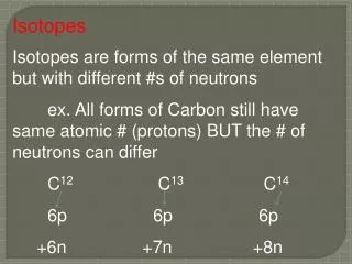
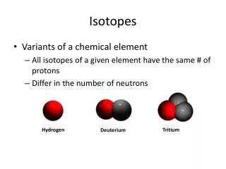
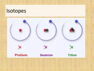
DEFINATION • Isotopes are nuclides with the same atomic number but different mass numbers . • The spontaneous degradation of nucleus & transmission of one element to another with consequent emission of rays ( or ) particles is known as radioactivity .
TYPES OF RADIO ACTIVE DECAY • 1 )Decay by negatron emission , • 2 )Decay by positron emission , • 3 )Decay by α particle emission , • 4 )Decay by gamma rays emission , • 5 )Decay by X rays emission . • Decay by Negatron emission : When Neutron is converted to a Proton by the ejection of a negatively charged β particle called a Negatron ( β- ) is emitted . Neutron Proton + Negatron
contd • Negatron emission is very important to biochemist . • 3 H & 14 C are β emitters can be used to label any organic compound . • 35 S used to label methionine , to study protein synthesis. • 32 P , a powerful tool in molecular biology & used as a nucleic acid label . • β emitting isotopes are most suitable for autoradiography , particularly for cell & tissue localization experiment .
DECAY BY POSITRON EMISSION • When Proton is converted to Neutron a positively charged β particle known as positrons( β+) is emitted . Proton Neutron + Positron . • Positrons are extremely unstable , they dissipate their energy in interaction with electrons .
Contd • The mass & energy of 2 particles( positron & electron ) are converted to 2 γ (gamma) rays are emitted at 180° to each other . • Positrons are detected by the same instrument used to detect γ radiation . • Positron emission tomography used to identify active & inactive areas of brain .
DECAY BY α PARTICLE EMISSION • Isotopes of elements with high atomic numbers frequently decay by emitting α particle . • An α particle is a helium nucleus contains 2 protons & 2 neutrons (4He2 +). • α particles have high ionizing power, less penatrance & are extremely toxic . • Isotopes that decay by α particle emission are not frequently encountered in biological work .
ELECTRON CAPTURE • Proton captures an electron orbiting in the inner most shell . Proton + Electron Neutron + X rays • Proton becomes a Neutron & electromagnetic X rays is given out .
DECAY BY EMISSION OF γ RAYS • These γ rays result from a transformation in the nucleus of an atom ( in contrast to X rays emission ) & frequently accompany α & β particle emission . • Emission of γ radiation leads to leads to no change in atomic number or mass . • γ radiation has low ionizing power but high penetration .
HALF LIFE OF RADIOISOTOPES • Half life of radio isotope is the time period required for radionuclide to decay to one half the amount originally present . • t1/2 = 0.693/λ. • λ is decay constant , a characteristic of a given isotope decaying in unit time .
UNITS OF RADIOACTIVITY • Bequerel is the unit of radioactivity is defined as one disintegration per second (1 d. p. s. ). • Frequently used units are curie , defined as the quantity of radioactive material in which the number of nuclear disintegrations per second is same as the 1gm of radium ( 3.7 X 10 10 Bq ). • Specific activity is defined as disintegration rate per unit mass of radioactive atoms.
Detection & Measurement of Radioactivity • Various methods for measuring radioactivity • 1) Autoradiography , • 2) gas ionization detectors & • 3) fluorescent scintillation , are the basis to detect & measure radioactivity in clinical laboratory .
AUTORADIOGRAPHY • In autoradiography a photo graphic emulsion is used to visualize molecules labeled with a radioactive element . • The emulsion consists of a large number of silver halide crystals embedded in a solid phase such as gelatin .
Contd • As energy from radioactive material dissipated in the emulsion , the silver halide becomes negatively charged & is reduced to metallic silver. • Photographic developers are designed to show these silver grains as blackening of the film , & fixers remove any remaining silver halide .
contd • Techniques of autoradiography have become more important in molecular biology . • Weak β – emitting isotopes ( 3H ,14 C ,35 S) are most suitable for autoradiography , particularly for cell & tissue localization experiments . • Low energy of negatrons & short ionizing track of isotope will result in discrete image .
contd • β emitting radioisotopes are used when radioactivity associated with subcellular organelles is being located . • 3H is the best radioisotope , since it’s all energy will get dissipated in the emulsion . • Electron microscopy can then be used to locate the image in the developed film .
contd • For location of DNA bands in electrophoretic gel, 32 P labeled nucleic acid probes are useful . • After hybridization ,hydrolysis & separation of DNA fragments by electrophoresis , a photographic plate is applied to to the covered gel & allowed to incubate .
Choice of emulsion & film • X ray films are generally suitable for macroscopic samples such as whole body, electrophoretographs , chromatographs . • When light (or) electron microscopic , detection of image ( cellular , subcellular localization of radioactivity ) very sensitive films are necessary .
contd • Time of exposure & film processing depends upon the isotope , sample type , level of activity , film type & purpose of the experiment. • In Direct autoradiography , the X ray film or emulsion is placed as close as possible to the sample .
Fluorography • Fluorography is used to cut short the time of exposure . • A fluorescent material such as ( PPO or sodium silicate )is infiltrated into the gel . • Negatrons emitted will excite fluorescent material & emit light , which will react with the film .
Intensifying screens • When 32 P labeled or γ –isotope labeled samples are used because of more penetrating nature of γ rays poor image is formed . • Intensifying screens helpful in giving a good image . • Solid phosphorus is applied on the other side of the film from the sample .
Other methods for amplifying image • Sensitivity of film is increased by preflashing . • Preflashing involves millisecond light flash prior to sample is being brought to juxtaposition with the film . • Low temperature exposure will provide higher sensivity.
GAS FILLED DETECTORS • Detectors filled with gases or gas mixtures are designed to capture & measure the ions produced by radiation within the detector ( excitation & ionization produces a pulse of current ). • Gas filled detectors used to measure radioactivity include 1) Ionisation chamber 2) Proportional counter 3) Geiger Muller counter • Geiger muller counter is used in clinical laboratory .
SCINTILLATION COUNTING • In scintillation process the radiation causes excitation & ionization of fluorescent material , the absorbed energy produces a flash of light. • The principal types of scintillation detectors found in clinical laboratory are 1) sodium iodide crystal scintillation detector . 2) the organic liquid scintillation detector.
APPLICATIONS OF RADIOISOTOPES IN BIOLOGICAL SCIENCES • Radioisotopes are frequently used for tracing metabolic path ways . • Mixing radiolabeled substrates & samples of the experimental material & collecting samples at various times , extract & separate the products by chromatography. • Radioactivity detectors can be attached to gas liquid chromatography or HPLC columns to monitor radioactivity coming off the column during separation .
uses • It is possible to predict the fate of individual carbon atoms of (14 C ) acetate through TCA cycle. • Methods have been developed to isolate intermediates of the cycle & to ascertain the distribution of carbon atoms within each intermediate( this is called as specific labeling pattern ) .
uses • Radioisotopes are used in ascertaining the turnover times for particular compounds . • Group of rats injected with radio labeled amino acid left for 24 hours allowing to assimilate into proteins. • The rats are killed at suitable time intervals & radioactivity in organs or tissue of interest is determined .
uses • Radioisotopes are widely used in study of the mechanism & rate of absorption , accumulation & translocation of inorganic & organic compounds in the animal . • Radiolabeled drugs are useful in pharmokinetic studies ( site of accumulation , rate of accumulation , rate of metabolism & metabolic products ) .
ANALYTICAL APPLICATIONS OF RADIOISOTOPES • Virtually any enzyme reaction can be assayed using radioactive tracer methods. • Radioisotopes have been used in study of 1) The mechanism of enzyme action & 2)In studies of ligand binding to membrane receptors.
contd • Isotope dilution analysis : when a known amount of radioactive tracer is introduced into an unknown volume , after thorough mixing , the concentration of radio tracer is estimated . V = N / n V = volume to be measured N = total number of counts injected n = number of counts per ml
Contd • By isotope dilution analysis plasma volume , total body water , E.C.F volume , RBC cell volume , total exchangeable sodium can be measured . • 131 I labeled human serum albumin useful in diagnosing protein losing enteropathy . • 51 Cr labeled RBC are given intra venously if there is any GI blood loss radioactivity can be measured .
Contd • Radio immuno assays are useful in analysis of hormones , growth factors , tumour markers , cytokines , bacterial antigens ,vitamin D & various biological molecules . • In RIA either antigen or antibody is radiolabeled . • Radiolabelling must not interfer in the binding of antigen & antibody , has to be compared with unlabeled ones .
Radioisotopes used in Diagnostic purposes • Radio active iodine uptake & imaging reveals the functional status of thyroid tissue , including nodules , the whole thyroid gland & metastatic foci . • 131I is used for thyroid cancer imaging & management . • 123 I is used for thyroid scan .
contd • Schilling test : used to detect the malabsorption of vitamin B12 . • Measurement of urinary radio labeled B12 following a saturation dose of non labeled stable B12 • 1000µg of non labeled B12 is given IM. • 1µg of labeled B12 is given orally. • Less than 5% excretion of radio labeled dose indicates malabsorption of Vit B12.
Contd • Technetium 99 m ( 99 m Tc ) pertechnetate: it is trapped by the thyroid gland but not organified , it can give a reasonable thyroid image even if patient is taking thyroid replacement therapy . • 99m Tc – MIBI ( 2 – methoxy 2 – methyl propyl isonitrile ) used in preoperative localization of parathyroid gland .
contd • Thalium 201 facilitates detection of 131 I negative metastatic thyroid cancer lesions in total body scan . • Iodo cholesterol 131I labeled 6 iodo methyl -19 norcholesterol , NP-59 used in adrenocortical imaging in cushing disease, cortisol producing adenoma , primary aldosteronism .
contd • MIBG ( 131 I or 123 I –meta iodo benzyl guanidine )scan is useful in adrenomedullary imaging in pheochromocytoma , neural crest tumors , carcinoid , medullary carcinoma thyroid . • Isotope bone scan is extremely useful in pagets disease of bone .
contd • Indium 111 octreotide scan a somatostatin analogue used to show : neural crest tumors, pheochromocytoma , carcinoid , paraganglioma & medullary carcinoma thyroid .
Contd • Fluorodeoxy glucose PET helpful in detection of 131 I negative thyroid carcinoma ,& MIBG negative pheochromocytoma . • Strontium 89 & Samarium 153 are two radionuclides that are preferentially taken in bone , particularly sites of new bone formation, capable of controlling bone metastasis .
contd • Xenon 133 is useful in lung function tests & is useful in diagnosing malfunctions of lung ventilation . • (133 I) iodohippuric acid used in diagnosis of kidney infections , kidney blockages or imbalance of function between two kidneys .
Contd • 51Cr –EDTA , 99m Tc-DTPA & 125I –iothalamate have clearance closest to inulin . ( useful in measurement of GFR ) • 99m Tc-DTPA has the advantage that it can also be used for gamma camera imaging .
Therapeutic uses of radioisotopes • Radioisotopes have role in management of malignancies . • Tumour tissues are attacked by beam of radiation . • 131I is used for treatment of thyroid cancer . • Teletherapy : 60Co is the source of radiation , radiation occurs from a distant source . • Radioactive material is impregnated into body in form of beeds or needles oe either as surface applicants .
contd • 60Co or radium rods are used in treatment of cervical cancer . • 32 P surafce applicants have role in Rx of squamous cell carcinoma , superficial angiomas , mycosis fungoides . • Boron 10 neutron irradiation has been recently used in the treatment of the inoperable & rapidly fatal brain tumour like glioblastoma multiforme
Contd • 48Au ( gold ) is used for treatment of malignant pleural & peritoneal effusions. • Yttrium90 synovectomy is useful in management of arthrites in hemophelics .
Radiation hazards • Immediate effects : 1 ) Bone marrow syndrome, 2 ) Gastrointestinal track syndrome, 3 ) Central nervous system syndrome . • Bone marrow syndrome : severe damage to hematopoietic system , leads to pancytopenia occurs with exposure of 200-1000 rads.
contd • Gastro intestinal syndrome : Severe damage to mucosal epithelium . Exposure of 1000 – 5000 rads is the cause . • Central nervous system syndrome : Blood brain barrier is lost . Exposure of 5000 – 10000 rads is the cause . • Delayed effects : carcinogenesis by damaging DNA
Radiation safety & protection • The most popular triad of radiation protection is time ,distance & shield (TDS). • Minimum possible time should spent near the radiation zone . • Handling of radioactive material should be done from maximum possible distance . • Person should be shielded by lead .