Download

1 / 21
210 likes | 412 Vues
Acute glomerulonephritis. Asist. Prof. Magdalena Stârcea IV th Pediatri c Clinic.

E N D
Acute glomerulonephritis Asist. Prof. Magdalena Stârcea IVth Pediatric Clinic
DEFINITION: condition characterized by inflammation of the renal glomeruli, often induced by aimmune trigger. Local inflammation and tissue proliferation of glomerular basement membrane damage may result in the glomerular mesangium and renal capillary endothelium, resulting in a decrease in the glomerular filtration rate and sodium retention of water. In acute glomerulonephritis patients have signs and symptoms such as hematuria, proteinuria, edema, hypertension. Glomerulonephritis may occur within kidney, or as a result of systemic diseases. Hippocrates originally described the manifestation of back pain and hematuria, which lead to oliguria or anuria. With the development of the microscope, Langhans was able to describe the corresponding glomerular changes.
ETIOLOGY: - 80% of cases - beta hemolytic streptococcus group A - bacteria (pneumococci, staphylococci, mycobacteria, Salmonella, Treponema pallidum, actinobacili) - viruses (virus Coxakie B, Echovirus type 9, influenza virus, measles virus, Cytomegalovirus, Coxsakie virus, Epstein Barr virus, hepatitis B virus) - parasites (Plasmodium malariae, Plasmodium falciparum, Schistosoma, Toxoplasma) - rickettsiae, fungi - systemic causes (Wegener's granulomatosis, systemic lupus erythematosus, cryoglobulinemia, polyarteritis nodosa, Henoch Schonlein) - systemic treatment with penicillin, gold compounds - primary glomerular disease (membranoproliferative glomerulonephritis, Berger's disease, mesangial proliferative glomerulonephritis pure)
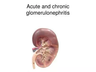
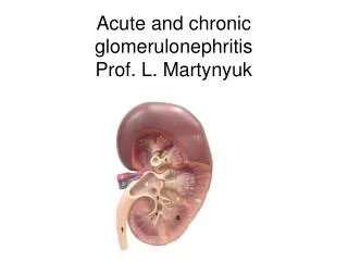
MORPHOPATHOPHY - Macroscopic: kidney enlarged, pale, with cotic kidney thicken. On gross appearance, the kidneys may be enlarged up to 50%.
Microscopy Optical- diffuse damage, increased glomerular volume bundle, capillary lumens compressed, glomerular cell proliferation and inflammatory infiltrators (monocytes, neutrophils, eosinophils). MBG is not uniformly thickened; sometimes cell proliferation capsule form crescents; normal renal tubules.
Electron Microscopy: dense deposits (humps) on the epithelial side of the GBM.
- Immunofluorescence reveals deposition of immunoglobulins and complement with fine, granular aspect ("starry sky")
Clinico-morphological correlations Low correlation between the severity of histological lesions and magnitude of proteinuria / haematuria Good correlation between glomerular cellularity and: decrease of GRF size of subendothelial deposits severity of the clinicalaspect
PATHOGENESIS: After streptococcal infection circulating immune complexes formed in excess deposit on GBM. At the same time consumption of complement is produced, so that the level of total serum complement decreases. Characteristic glomerular lesions are the result of in situ formation and deposit of immune complexes. As a result of the presence of these deposits glomeruli appear edematous and inflammatory cell infiltrates. In poststreptoccocical glomerulonephritis triggers are derived from streptococcal protein. The decrease in glomerular filtration causes oliguria, hematuria, proteinuria, impaired glomerular filter by, the activation of the renin-angiotensin - aldosterone system with hypertension, ADH activation by increasing the circulating volume swelling.
History: • The typical patient is a boy, aged 2-14 years, who suddenly develops puffiness of the eyelids and facial edema. The urine is dark and scanty; the blood pressure is elevated. • Symptom onset usually is abrupt.Nonspecific symptoms include weakness, fever, abdominal pain, and malaise. • In the setting of postinfectious acute nephritis, a latent period of up to 3 weeks occurs before onset of symptoms. • The latent period generally is 1-2 weeks for the postpharyngitis form of the disease and 2-4 weeks in the case of postdermal infection. • Onset of nephritis within 1-4 days of streptococcal infection suggests preexisting renal disease.
Patient evaluation should include: • Anthropometric data: weight, height, body surface area • Monitorizing of BP (every 3 hours) • Evaluation of edematous syndrome (peripheral, ascites, pleural effusion) • Evaluating cardiovascular system: signs of fluid overload (tachycardia, congestive hepatomegaly, hepato-jugular reflux, respiratory failure) • 24-hour urine volume measurement, metering ingestion / diuresis
Latency period: 3 weeks after streptococcal throat infection (3-6 weeks of streptococcal infection skin) and is asymptomatic. • Period status: - Edema - peripherals to anasarca, depending on the severity of renal damage, the amount of liquid ingested and extent of proteinuria - Macroscopic haematuria: half of patients, the urine aspect of "Douche of flesh" that lasts up to 4 weeks -Hypertension: moderate to severe value well tolerated in children; in 5% of cases may be complicated by hypertensive encephalopathy; sometimes complicated by pulmonary edema (dyspnea with orthopnea, rales crepitation rises from the bottom, dry cough, respiratory failure) - Oliguria (less 1ml/kg/hour) - Proteinurialess than 50mg/kg/dailly (nephritic level) - Signs of systemic disease: malar rash, arthritis / arthralgia, purpura, vascular, etc.
LABORATORY STUDIES: 1. Urinanalysis - Reduced urinary volume - Increased urinary density > 1020 - Osmolarity as 700mOsm / l - Proteinuria in 50mg/kg/day or below 2 g / m² / day ( disappearswithin 6 months) - Hematuria (most constant sign ) - disappears after more than 1 year ( maintaining its accompanying proteinuria after 1 year is a sign of chronic glomerular damage ) with dysmorphic red blood cells (the fresh urine specimen) - Hematic cats (Addis test , urinary sediment)
2. SerologicalExplorations - Exploring arranged function Renal urea , creatinine , creatinine clearance , GFR ( Schwartz formula ) - Blood electrolytes : hyperkalemia , metabolic acidosis - CBC : anemia ( by hemodilution ) - Inflammatory Syndrome : ESR, CRP , fibrinogen - Hypoproteinemia by hemodilution rare type of losses due to nephrotic 3. ImmunologicalExplorations - Decreased total serum complement , fractions C3 , C4 , C5 (normalized within 8 weeks ) - The presence of type III cryoglobulins (IgM , IgG and C3) • Specific determinations of systemic diseases : ANA , ds DNA Ac 4. Bacteriological Explorations :- ASO , anti DNA - za B Ac Ac antihyaluronidase- Pharyngeal cultures or skin lesions
5. Imaging studies - Chest radiograph : in patients with chronic cough with orwithout hemoptysis (APE) - Echocardiography , ECG : evaluation of hypertensive heart , highlighting endocarditis , pericarditis arrhythmias - Renal ultrasound : a process dimension diagnosed chronic kidneyshowed low acute - Cranio- cerebral CT in children with altered mental status , HIC phenomena , focal deficits installed under malignant hypertension
Kidney biopsy indications: - Absence of infection before the onset- Intrainfectious AGN - Normal level of complement - Presence of impure nephrotic syndrome- Azotemia without other clinical signs- Age under 2 years- History of kidney disease- Systemic symptoms ( prolonged fever, arthralgia / arthritis, liver disease, hemtologic disease, rash)- Oliguria / azotemia more than 2 weeks- HTA more than 3 weeks- C3 lower more than 8 weeks- Proteinuria and hematuria more than 6 months- Microscopic haematuria over 1 year
TREATMENT: • Antibiotic therapy : not indicated.Benzatilpenicilina G may be recommended at a dosage of 100 000 IU / kg / day in four divided doses for 10 days, only if it is initiated early after the onset of streptococcal disease. Can sterilize any remaining focars, but no prevents AGN! • Antihypertensive therapy : • hipertensive encephalopathy :- Diazoxide iv bolus 5mg/kg/dosis - Na nitroprusside : 0.5 - 8μg/kg/min , iv- Furosemide: 1 - 2mg/kg , iv slow • severe hypertension without encephalopathy :- Minoxidil 0,1 - 0,2 mg / k g / day , max 5mg/24 hours- Ca channel blockers : from 0.25 to 0.5 mg / kg / dose , max 1-2mg/kg/zi Warning: angiotensin converting enzyme inhibitors may further reduce glomerular filtration and beta-blockers may exacerbate hyperkalemia ! • acute pulmonary edema : hemodyalisis
3. Supportive Treatment is aimed disorderselectrolytic , especially hyperkalemia : • K < 6.0 mmol / l- stop the K sparing therapy, food restriction (restriction dried fruit, bananas, citrus fruits, beans, dried peas, green vegetables, potatoes) • K - 6.0 - 6.5 mmol / l :- hydration- Slow bolus Ca gluconate 10% , 0.5 ml / kg, ECG monitoring- PIV SG 10-30 % 1ml/kg ( rapid insulin 1U/5g glucose)- Sorbitol 70 % + Kayexalate the 1g/kgc/po , 2ml/kg- Where AR < 18 mmol / l - 8.4 % Na bicarbonate , 2mEq/kg/day in 4 parts- K repeated after 6 hours • K > 6.5 mmol / l :- Continuous ECG monitoring- Salbutamol nebuliser- Emergency hemodialysis
4. Hygienic-dietary regime :- Daily weighing , measuring from 3:00 to AP- The oligo - anuria below 0.5 ml / kg / h - fluid restriction ( diuresis + 400 ml )- Restriction of salt, protein restriction in the case of azotemia 5. Hemodialysis/hemofiltration/peritoneal dialysis - Heart failure, pulmonary edema- Hypertensive encephalopathy refractory to therapy- Severe hyperkalemia , rebel seizure therapy- Secondary uremic gastritis .
References: 1. Mihaela Munteanu, Glomerulonefritele acute (postinfecțioase), în Hematologie și nefrologie pediatrică, elemente practice de diagnostic și tratament, editura Junimea, Iași, 2008, cap. 6, pag. 239 – 249. 2. KDIGO Clinical Practice Guideline for Glomerulonephrites, Chapter 9: Infection-related glomerulonephritis, Kidney International Supplements (2012) 2, 200 - 208; 3. Nelson textbook of pediatrics - 18 ed. Behrman RE, Kliegman RM, Jenson HB: Glomerulonephrites associated with Infections, cap. 511, pag. 2173 – 2175, Saunders Elsevier, 2007. 4. Patrick H. Nachman, J. Charles Jennette, Ronald J. Falk, Primary Glomerular Disease, cap. 31, pag. 1100 – 1165, în BRENNER & RECTOR’S THE KIDNEY, ediția 9, , Saunders Elsevier, 2012. 5. Rees L., Brogan P., Bockenhauer D., Webb N., Glomerular disease, în Pediatric Nephrology, Oxford Specialist Handbooks in Pediatrics, second edition, cap. 9, pag. 181 – 224.