Download

1 / 39
720 likes | 1.92k Vues
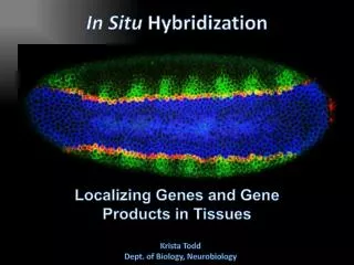
In Situ Hybridization. Localizing Genes and Gene Products in Tissues. Krista Todd Dept. of Biology, Neurobiology. Monitoring Nucleic Acids. DNA RNA Protein . Determine the chromosomal location of a gene

E N D
In Situ Hybridization Localizing Genes and Gene Products in Tissues Krista Todd Dept. of Biology, Neurobiology
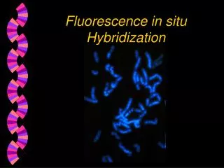
Monitoring Nucleic Acids DNA RNA Protein • Determine the chromosomal location of a gene • FISH (Fluorescence in situ hybridization)
F I S HFluorescence In Situ Hybridization S. Mewborn, U Chicago
Monitoring Nucleic Acids DNA RNA Protein • Determine the chromosomal location of a gene • FISH (Fluorescence in situ hybridization) • Monitor amount an identity of currently translated RNAs • qRT-PCR, TRAP (translating ribosome affinity purification)
T R A P(translating ribosome affinity purification) Cell November 2008
Monitoring Nucleic Acids DNA RNA Protein • Determine the chromosomal location of a gene • FISH (Fluorescence in situ hybridization) • Monitor amount of currently translated RNAs • qRT-PCR, TRAP (translating ribosome affinity purification • Monitor mRNA amount, location and experimentally-induced changes/kinetics • Laser-capture micro-dissection/microscopy • Whole mount In situ hybridization
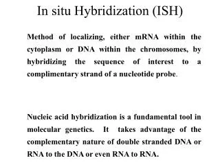
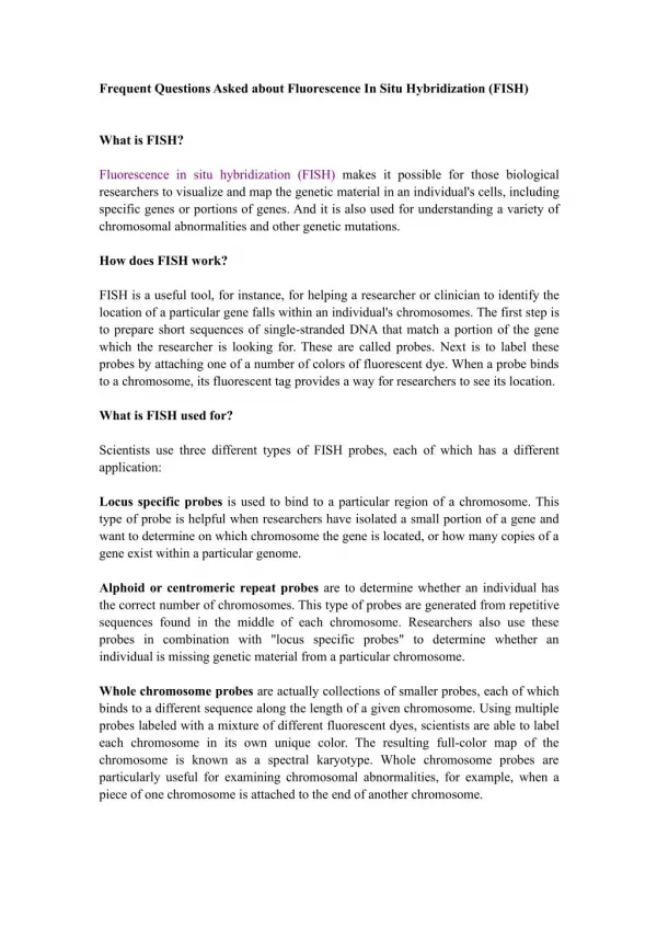
In situ Hybridization Method of localizing, either mRNA within the cytoplasm or DNA within the chromosomes, by hybridizing the sequence of interest to a complimentary strand of a nucleotide probe. Creative Biolabs
When/Why do in situ hybridizations • Distribution of protein and RNA are different • Protein transport, extracellular/secreted proteins • Different temporal changes in protein and RNA • Analysis of whole tissue might not be sensitive enough • Single cell sensitivity • RNA is not uniformly distributed in a cell • e.g. neurons: axon vs. soma vs. dendrite • No antibody available
In situ Hybridization:Applications • Quantitative methods and Qualitative methods • in situ hybridization to metaphase chromosome spreads • RNA detection by in situ hybridization • Whole mount hybridization • In situ hybridization in electron microscopy • Valuable control for RNAi experiments
In situ Hybridization:Applications • Quantitative methods and Qualitative methods • in situ hybridization to metaphase chromosome spreads • RNA detection by in situ hybridization • Whole mount hybridization • In situ hybridization in electron microscopy • Valuable control for RNAi experiments
Principle Methods:In Situ Hybridization for microscopy • Probes • Types • Labeling • Tissue Preparation • Probe Detection
Probes • Oligonucleotides: • single stranded DNA (RNase resistant) • Short 20-50 bases (good tissue penetration) • Cover only part of the mRNA, but potentially highly specific • Single stranded DNA (200-600 bases) • Produced by Reverse transcription of RNA or primer amplified • Double stranded DNA • denaturation necessary • only one strand is specific • Less sensitive due to self hybridization • RNA • RNA-RNA hybrids are very stable and RNase resistant • Post hybridization digestion with RNase possible Hybrid Bond Strength RNA-RNA > RNA-DNA > DNA-DNA
Probe Labeling • Non-radioactive • Biotin • Digoxigenin • Fluorophore
Labeled nucleotides • Digoxigenin • Biotin • Fluorophore
Probe Labeling • Non-radioactive • Biotin • Digoxigenin • Fluorophore • Radioactive Advantage: sensitivity Disadvantage: hazard, long exposure times S35; P33; P32;H3
Probe labeling • Biochemical Synthesis with labeled nucleotides • Terminal Labeling: 3’ and 5’ ends • Nick translation • Random primed labeling • PCR • In vitro transcription
32P 5’OH 32P 5’OH DNA Terminal Labeling • 3’ terminals: tailing Deoxynucleotidyltransferase catalyzes repetitive addition of mononucleotides from a dNTP to the terminal 3’OH • 5’ terminals: T-4 polynucleotide kinase transfers the gamma-phosphate residue to the terminal 5’ phosphate group of a DNA molecule g32P-ATP & POLYNUCLEOTIDE KINASE
DNA Probe Labeling • Nick translation: • DNaseI + DNA-polymerase + labeled dNTP 3’ 5’ 5’ 3’ 5’ 3’ 5’ 3’ 5’ 3’ 5’ 3’
In vitro Transcription of RNA probe Antisense: Cut with EcoRI Use T-3 polymerase EcoRI T7 BamHI T3 Sense: Cut with BamHI Use T-7 polymerase
Post-synthesis labeling Psoralen biotin
Advantages of RNA probes: • RNA-RNA hybrids are very stable • Tissue can be digested with RNase (dsRNA is not digested) after the hybridization reducing the background • Higher specific activity compared to oligonucleotides • Strand-specific compared to dsDNA probes • Advantages of oligonucleotide probes: • Better tissue penetration • Multiple oligos can be mixed to enhance signal
Tissue Procedures • Tissue Fixation • Permeabilization • Pre-Hybridization • Hybridization • Development and Visualization
Tissue Preparation • Detergents: Triton, SDS (permeabilization) • Proteinase K (permeabilization) • Enzyme neutralization: H2O2 for peroxidase, levamisole for alkaline phosphatase • Acetylation: 0.25 % acetic anhydride in triethanolamine(neutralization of positive charges) • HCl (protein extraction and denaturation of target sequence)
Effect of Fixation and Proteinase Digestion 4% paraformaldehyde 2.5 % glutaraldehyde 0.05% 0.02% 0.005% 0.002% Proteinase K Spinal Cord; probe PLP mRNA
Standard hybridization buffer • Buffer: Tris or phosphate • 2xSSC: NaCl/sodium citrate: monovalent cations • Formamide: reduces thermal stability/TM • EDTA (1mM) chelator to remove divalent cations, which strongly stabilize DNA/RNA hybrids • Yeast tRNA (500 µg/ml): blocking agent • Dextran sulfate (10%) reduce free volume and therefore probe concentration
Factors Influencing Hybridization • Temperature • Salt concentrations: Na+ • Organic solvents: e.g. formamide • Probe length • G/C content (<55-60%) • Volume (dextran sulfate)
Melting Temperature of Nucleic Acids • TM = 81.5˚C+16.6(logM)+0.41(%GC)-500/n M = salt molarity / n=length of probe • Probe: • G/C content • Length of probe • Higher Stringency of washes: • Higher temperature • Lower salt concentration • Higher formamide concentration
Signal Visualization • Radioactive: film/emulsion • Fluorescence • Enzymatic reaction: AP, HRP • Signal Amplification
Multiplex mRNA detectionDave Kosman (Ethan Bier and Bill McGinnis labs, UC San Diego) http://superfly.ucsd.edu/%7Edavek/images/quad.html
Tyramide Signal Amplification (TSA) Up to 100x signal amplification
Controls • Specificity of probe • Sequence analysis • Testing by Northern blot • Negative controls: • RNase treatment pre-hybridization • Addition of an excess of unlabeled probe • Hybridization with sense probe • Tissue known not to express the gene of interest • Positive Controls: • Comparison with protein product • Comparison to probes hybridizing to different part of the same mRNA • Tissue known to express the gene of interest • Poly dT probe or housekeeping gene to check RNA integrity
Quantification • Measurement of relative RNA amounts: • Assumption: signal intensity is proportional to the amount of cellular RNA, labeling is within the linear range of the emulsion and quantification system • Difficulties: • Comparison of different probes • Comparison of different runs • Influences of storage, temperature, probe concentration, tissue thickness, emulsion thickness, development times • Cellular Level: • Density measurements • Count silver grains • Tissue Level:densitometry on x-ray film