
Absence of PKH-Stained Liposomes in Injured Spinal Cord Post-Injection
This study investigates the uptake of PKH-stained liposomes in the spinal cord following injury. Brightfield microscopy revealed control unstained PBS liposomes (A), while no fluorescence was observed (C). PBS liposomes were effectively stained with PKH26, indicated by red fluorescence (D). Comparison of spleens showed EGFP+ fluorescence in control mice (E) without PKH26 positivity (G). Interestingly, one day after spinal cord injury (SCI), mice injected with PKH26-stained liposomes exhibited both EGFP+ and PKH26+ fluorescence (F, H), yet no PKH26+ liposomes were located at the lesion epicenter (I-K, L-M, O-Q).

Absence of PKH-Stained Liposomes in Injured Spinal Cord Post-Injection
E N D
Presentation Transcript
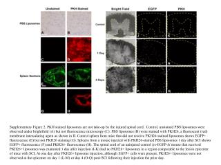
Supplementary Figure 2. PKH stained liposomes are not take-up by the injured spinal cord. Control, unstained PBS liposomes were observed under brightfield (A) but not fluorescence microscopy (C). PBS liposomes (B) were stained with PKH26, a fluorescent (red) membrane intercalating agent as shown in D. Control spleen from mice that did not receive PKH26-stained liposomes shows EGFP+ fluorescence (E) but not PKH26 staining (G). Spleens from a mouse injected with PKH26-stained PBS liposomes 1 day after SCI shows EGFP+ fluorescence (F) and PKH26+ fluorescence (H). The spinal cord of an uninjured control lys-EGFP-ki mouse that received PKH26+ liposomes was examined 1 day after injection (I-K) had no PKH26+ liposomes in a region comparable to the lesion epicenter of mice with SCI. At one day after PKH26+ liposome injection, although EGFP+ cells were present, PKH26+ liposomes were not observed at the epicenter on day 1 (L-M) or day 4 (O-Q) post-SCI following their injection the prior day.