Download
1 / 63
640 likes | 765 Vues
Mechanisms of Disease: histone modifications in Huntington's disease Ghazaleh Sadri- Vakili and Jang-Ho J Cha* Nature Clinical Practice Neurology (2006) 2, 330-338.

E N D
Mechanisms of Disease: histone modifications in Huntington's diseaseGhazaleh Sadri- Vakili and Jang-Ho J Cha* Nature Clinical Practice Neurology (2006) 2, 330-338
In 1872, the American physician George Huntington wrote about an illness that he called "an heirloom from generations away back in the dim past." and described the disorder in his first paper "On Chorea" at the age of 22 "In the history of medicine, there are few instances in which a disease has been more accurately, more graphically or more briefly described."
One of its earliest names was chorea, which, as in "choreography," is the Greek word for dance. The term chorea describes how people affected with the disorder twist, and turn in a constant, uncontrollable dance--like motion. • "Hereditary chorea" emphasizes how the disease is passed from parent to child. • "Chronic progressive chorea" stresses how symptoms of the disease worsen over time. • Today, physicians commonly use the simple term Huntington's disease (HD) to describe this highly complex disorder that causes untold suffering for thousands of families.
Examining the combined medical history of several generations of a family exhibiting similar symptoms, he realized their conditions must be linked; he presented his detailed and accurate definition of the disease as his first paper. Unknowingly, described the exact pattern of inheritance of autosomal dominant disease years before the rediscovery of Mendelian inheritance • During the rediscovery of Mendelian inheritance at the turn of the 20th century, HD was used tentatively as an example of autosomal dominant inheritance
Huntingtin (Htt), the protein that is defective in HD, is expressed throughout the body, principally affects the brain. • including the cortex, thalamus and sub thalamic nucleus, the striatum is the most severely affected region. • In striatum, the medium spiny projection neurons are preferentially targeted by the disease and interneurons are relatively spared • preferential loss of medium spiny neurons and sparing of interneuron populations indicates that neurodegeneration in HD is cell-type-specific.
Htt is expressed in all mammalian cells. • The highest concentrations are found in the brain and testes • The function of Htt in humans is unclear. • It interacts with proteins which are involved in transcription, cell signaling and intracellular transporting. • In animals genetically modified to exhibit HD, several functions of Htt have been found. • In these animals, Htt is important for embryonic development, as its absence is related to embryonic death. • It also acts as an anti-apoptotic agent preventing programmed cell death and controls the production of brain-derived neurotrophic factor, a protein which protects neurons and regulates their creation during neurogenesis.
Htt also facilitates vesicular transport and synaptic transmission and controls neuronal gene transcription. • If the expression of Htt is increased and more Htt produced, brain cell survival is improved and the effects of mHtt are reduced, whereas when the expression of Htt is reduced, the resulting characteristics are more typical of the presence of mHtt. • In humans the disruption of the normal gene does not cause the disease. • It is currently concluded that the disease is not caused by inadequate production of Htt, but by a gain of toxic function of mHtt.
THE HUNTINGTON'S DISEASE MUTATION • is caused by a mutation in the IT15 gene on the short arm of chromosome 4. • This gene, which was renamed HD, consists of 67 exons that encode Htt, a 350-kD protein of 3,144 amino acids. • mutation is an expansion of the cytosine–adenine–guanine (CAG) trinucleotide repeat in exon 1, which codes for a pg moiety in the Htt protein. • Normal individuals have CAG repeat lengths of 7–34. • CAG repeat is expanded and unstable in HD patients • Repeat lengths of more than 40 glutamines produce HD, and repeats of over 70 glutamines invariably cause juvenile onset.
When the HD mutation was eventually identified as a CAG trinucleotide repeat expansion, HD joined a novel class of neurodegenerative disease, the polyglutamine diseases(PGD) • expansion of the CAG repeat occurs within the coding region of the gene, so the CAG repeat is translated into a polyglutamine stretch • PGD are all autosomal dominant or sex-linked dominant diseases • In both humans and transgenic mouse models, these disorders are characterized pathologically by pg protein-containing intracellular inclusions • the pg moiety is strongly implicated as the portion of the protein that makes the greatest contribution to disease pathogenesis.
As the disease progresses, concentration on intellectual tasks becomes increasingly difficult. • the disease may begin with uncontrolled movements in the fingers, feet, face,…. • The disease can reach the point where speech is slurred and vital functions, such as swallowing, eating, speaking, and especially walking, continue to decline. Some individuals cannot recognize other family members • The most common causes of death are infection (most often pneumonia), injuries related to a fall, or other complications
Huntington's disease has autosomal dominant inheritance, meaning that an affected individual typically inherits one copy of the gene with an expanded trinucleotide repeat (the mutant allele) from an affected parent. • In this type of inheritance pattern, each offspring of an affected individual has a 50% risk of inheriting the mutant allele and therefore being affected with the disorder . This probability is sex-independent.
Is there any age limit………………….. • Some individuals develop symptoms of HD when they are very young--before age 20.--"early-onset" or "juvenile" HD • and death often follows within 10 years. • A few individuals develop HD after age 55. • Diagnosis in these people can be very difficult. • The symptoms of HD may be masked by other health problems, or the person may not display the severity of symptoms seen in individuals with HD of earlier onset.
A baby History • To verify the link between the number of CAG repeats in the HD gene and the age at onset of symptoms, scientists studied a boy who developed HD symptoms at the age of two, one of the youngest and most severe cases ever recorded. • They found that he had the largest number of CAG repeats of anyone studied so far--nearly 100. • The boy's case was central to the identification of the HD gene and at the same time helped confirm that juveniles with HD have the longest segments of CAG repeats, the only proven correlation between repeat length and age at onset.
Heart Breaking or Interesting……? • The great American folk singer and composer Woody Guthrie died on October 3, 1967, after suffering from HD for 13 years. • He had been misdiagnosed, considered an alcoholic, and shuttled in and out of mental institutions and hospitals for years before being properly diagnosed. • His case, sadly, is not extraordinary, although the diagnosis can be made easily by experienced neurologists
The physician may ask the individual to undergo a brain imaging test. Computed tomography (CT) and magnetic resonance imaging (MRI) provide excellent images of brain structures with little if any discomfort. • Those with HD may show shrinkage of some parts of the brain—particularly two areas known as the caudate nuclei and putamen—and enlargement of fluid-filled cavities within the brain called ventricles. These changes do not definitely indicate HD, • When used in conjunction with a family history and record of clinical symptoms, however, CT can be an important diagnostic tool. • Another technology for brain imaging includes positron emission tomography (PET,) which is important in HD research efforts but is not often needed for diagnosis.
The most prominent early effects are in a part of the basal ganglia called the neostriatum, which is composed of the caudate nucleus and putamen. • Other areas affected include the substantia nigra, layers of cerebral cortex, the hippocampus, purkinje cells in the cerebellum, lateral tuberal nuclei of the hypothalamus • reducing in size as they lose cells Striatal spiny neurons • The basal ganglia—the part of the brain most prominently affected by HD—play a key role in movement and behavior control. • current theories propose that they are part of the cognitive executive system and the motor circuit.
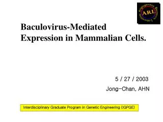
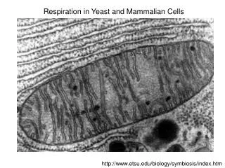
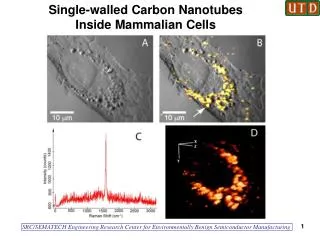
Area of the brain damaged by Huntington's disease – striatum (shown in purple)
The basal ganglia ordinarily inhibit a large number of circuits that generate specific movements. • To initiate a particular movement, the cerebral cortex sends a signal to the basal ganglia that causes the inhibition to be released. • Damage to the basal ganglia can cause the release or reinstatement of the inhibitions to be erratic and uncontrolled, which results in an uncontrolled movement • The accumulating damage to this area causes the characteristic erratic movements associated with HD.
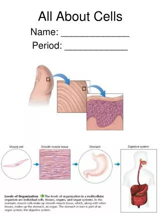
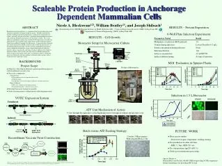
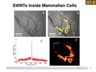
Coronalsection from a MRbrain scan of a patient with HD showing atrophy of the heads of the caudate nuclei, enlargement of the frontal horns of the lateral ventricles
How is Huntington's disease diagnosed? • The doctor will also ask about recent intellectual or emotional problems, which may be indications of HD, and will test the person's hearing, eye movements, strength, coordination, involuntary movements (chorea), sensation, reflexes, balance, movement, and mental status, and will probably order a number of laboratory tests as well. • People with HD commonly have impairments in the way the eye follows or fixes on a moving target
The discovery of the HD gene in 1993 resulted in a direct genetic test to make or confirm a diagnosis of HD in an individual who is exhibiting HD-like symptoms. • Using a blood sample, the genetic test analyzes DNA for the HD mutation by counting the number of repeats in the HD gene region.
A model for HD pathogenesis :pathogenic mechanisms • HD pathogenesis begins with altered conformation of the protein containing the expanded pg repeat. • Proteolysis generates a fragment that leads to toxicity through several pathways. • Nuclear importation may lead to altered gene transcription with a detrimental effect on cell survival. • Inclusions also form in the nucleus, but may not be a major cause of cell death.
Huntington fragments may interfere with mitochondrial energy metabolism, either directly, or more likely indirectly, perhaps via altered gene transcription. • Micro aggregation of the fragment may lead to caspase activation and the consequent initiation of cell death pathways. • Fragments may be transported into neuritis, interfering with cytoskeleton function.
Autophagy has emerged as a mechanism of HD pathogenesis mutant Htt accumulation activates the endosomal– lysosomal system and contributes to Htt proteolysis and autophagic cell death. • Inhibition of mTOR (mammalian target of rapamycin; also known as FRAP) activates autophagy and attenuates Htt-induced toxicity, indicating that autophagy might in fact be helpful as a clearance mechanism
There are multiple cellular changes through which the toxic function of mHtt may manifest and produce the HD pathology. • During the biological process of posttranslational modification of mHtt, cleavage of the protein can leave behind shorter fragments constituted of parts of the pg expansion. • The polar nature of glutamine causes interactions with other proteins when it is overabundant in Htt proteins. • Thus, the Htt molecule strands will form hydrogen bonds with one another, forming a protein aggregate rather than folding into functional proteins.
Over time, the aggregates accumulate, ultimately interfering with neuron function because these fragments can then misfold and coalesce, in a process called protein aggregation, to form inclusion bodies within cells. • Neuronal inclusions run indirect interference. • The excess protein aggregates clump together at axons and dendrites in neurons which mechanically stops the transmission of neurotransmitters because vesicles (filled with neurotransmitters) can no longer move through the cytoskeleton. • Ultimately, over time, less and less neurotransmitters are available for release in signaling other neurons as the neuronal inclusions grow. • Inclusion bodies have been found in both the cell nucleus and cytoplasm.
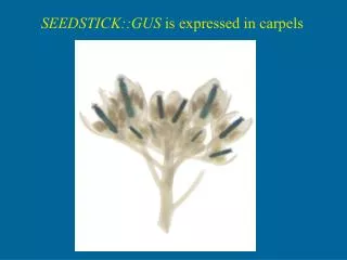
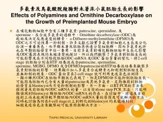
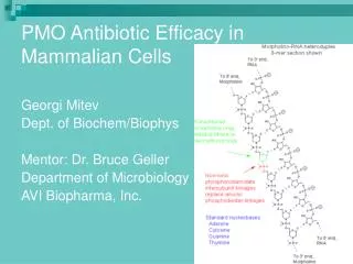
A microscope image of Medium spiny neurons (yellow) with nuclear inclusions (orange), which occur as part of the disease process
TRANSCRIPTIONAL DYSREGULATION IN HD • transcriptional dysregulation is an important underlying mechanism in HD pathogenesis. • Pg repeats in the N-terminal region of Htt protein gives it structural similarities to known transcription factors and repeat expansion leads to aberrant cleavage by caspases • The cleaved fragments gain access to the nucleus and form nuclear aggregates that might disrupt transcription • interacts with numerous transcription factors including CREB-binding protein (CBP), TATA-binding protein (TBP), p53 and Sp1 leads to Htt nuclear aggregation cause transcriptional dysregulation
Transcriptional dysregulation has been proposed to have an important role in the pathology of HD • control of eukaryotic gene expression depends on the modification of histone proteins associated with specific genes, with histone acetylation playing a crucial role. • Acetylation of histones at specific residues increases gene transcription; conversely, histone deacetylation represses transcription. Recent studies in numerous HD models have demonstrated a potential therapeutic role for histone deacetylase (HDAC) inhibitors in the treatment of polyglutamine diseases.
R6 mice were created by inserting exon 1 of the human HD gene containing 150 pg repeats, under the control of the human HD promoter, into the mouse genome. • Alterations of neurotransmitter receptor protein levels, as well as of mRNA levels, have been reported in the R6/2-before the onset of abnormal symptoms • In very early-grade HD cases there are profound reductions in the expression levels of cannabinoid CB1, dopamine D2 and adenosine A2a receptors. • screening of mRNA levels, using DNA microarrays, in the brains of R6/2 mice confirmed the finding that specific genes are downregulated in this HD mouse model.
It affects neuronal disfunction,neurotransmitter signaling….. • Gene expression studies have also been carried out in other HD mouse models. • The HD-N171-82Q transgenic mouse model expresses a COMPLEMENTARY DNA that encodes a 171-amino-acid N-terminal fragment of Htt exon 1 containing 82 CAG repeats. • The changes in gene expression in HD-N171-82Q mice are similar to those observed in R6/2 mice • changes are most pronounced in the striatum, with the motor cortex affected to a lesser degree, and the cerebellum showing the fewest gene changes. • Tm models expressing full-length versions of Htt, such as the YAC72 mice, demonstrate fewer gene changes, indicating that increasing the length of Htt reduces the severity of pg-induced gene changes
Recently, researchers have shown that mutant versions of N-terminal Htt can dissociate components of the transcriptional complex on gene promoters • transcriptional dysregulation is likely to be an important mechanism in the pathogenesis not only of HD, but also of other pg disorders
HISTONE MODIFICATIONS • The N-terminal tails of the core histones (H2A, H2B, H3 and H4) are strongly basic---contain specific amino-acid residues ---sites for several post-translational modifications, including acetylation, methylation, phosphorylation • acetylation of lys residues corresponds to transcriptionally active chromatin, whereas methylation of lys and arg residues leads to transcriptional repression. • acetylation of lys 9 and 14 on histone H3 correlates with active chromatin and leads to transcription, whereas methylation of lys 9 on histone H3 is a marker of silenced genes
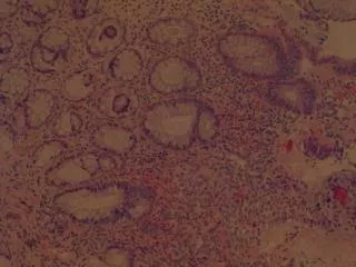
Histone acetylation and deacetylation are modulated by the interplay between histone acetyltransferases (HATs) and HDACs • HAT activity leads to increases in gene transcription by creating a more open conformation of chromatin, whereas HDACs remove acetyl groups, leading to gene repression through condensation of chromatin • Abnormalities of histone acetylation have been associated with a number of human cancers like leukemia non-Hodgkin's lymphoma • In human gastrointestinal cancers, histone acetylation is globally reduced
Acetylation of core histone proteins by the activity of HAT proteins or treatment with HDAC inhibitors ---more open conformation of chromatin---transcriptionally active state. Removal of acetyl groups by HDAC leads ----repressed chromatin----- transcriptional repression. Mutant Htt expression --- repressed chromatin acetylation of histones
HDAC inhibitors increase acetylation of histones, thereby increasing transcription of genes that have been silenced. • promote growth arrest by inducing the expression of tumor suppressor genes, and are commonly used as anticancer • Recent studies in yeast, cell culture, Drosophila and mouse models of pg disease indicate that HDAC inhibitors might be useful as therapeutic agents in HD
HISTONE DEACETYLASE INHIBITORS IN HUNTINGTON'S DISEASE • Many Htt-interacting proteins possess histone-modifying activity • CBP contains an acetyltransferase domain and is a coactivator at a number of promoters • overexpression of CBP decreases pg-induced cell death • Htt exon 1 with 51 glutamines containing the polyproline domain directly binds to the acetyltransferase domain of CBP and p300/CBP-associated factor • expression of Httex1p 20Q or Httex1p 103Q in PC12 cells causes a global hypoacetylation of histones, an effect that is reversed by the presence of the HDAC inhibitors sodium butyrate, trichostatin A (TSA) and suberoylanilide hydroxamic acid (SAHA)
in transgenic Drosophila that express Httex1p 93Q, a decrease in neuron degeneration and early adult death are observed if the flies have been reared on SAHA or sodium butyrate • These results indicated that reduced acetyltransferase activity might be an important component of pg pathogenesis in vivo, and paved the way for HDAC inhibitor studies in transgenic mouse models of HD. • Then investigated the effects of HDAC inhibitors in R6/2 mice. • Sodium butyrate treatment significantly prolonged the survival of R6/2 mice. • sodium butyrate and SAHA improved performance on a ROTAROD TEST, and were neuroprotective
there was a decrease in gross brain atrophy and ventricular enlargement in the sodium-butyrate-treated mice,and a reduction in neuronal atrophy in the striatum of SAHA-treated mice. • There was no effect on Htt aggregates • In both studies, administration of HDAC inhibitors corrected global hypoacetylation of histones. • In sodium-butyrate-treated R6/2 mice, there was an increase in Sp1 acetylation, but no change in basal levels of Sp1 was detected. • Sodium butyrate also provided protection against 3-nitropropionic-acid (3-NP)-induced striatal damage in R6/2 mice.