Chapter 5 Histology
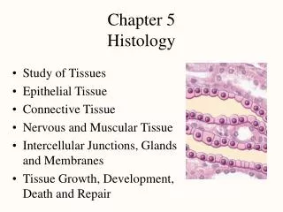
Chapter 5 Histology. Study of Tissues Epithelial Tissue Connective Tissue Nervous and Muscular Tissue Intercellular Junctions, Glands and Membranes Tissue Growth, Development, Death and Repair. The Study of Tissues. Whole body contains only 200 different cells types

Chapter 5 Histology
E N D
Presentation Transcript
Chapter 5Histology • Study of Tissues • Epithelial Tissue • Connective Tissue • Nervous and Muscular Tissue • Intercellular Junctions, Glands and Membranes • Tissue Growth, Development, Death and Repair
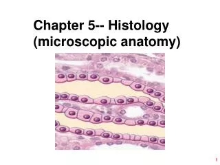
The Study of Tissues • Whole body contains only 200 different cells types • Four primary tissue classes • epithelial tissue • connective tissue • muscular tissue • nervous tissue • Histology (microscopic anatomy) • study of tissues and how they form organs • Organ = structure with discrete boundaries • composed of 2 or more tissue types
Features of Tissue Classes • Tissue = group of similar cells and cell products • arose from same region of embryo • Main differences between primary tissue classes • types and functions of cells • characteristics of the matrix (extracellular material) • fibrous proteins • ground substance • clear gel called many different names (ECF, tissue fluid, interstitial fluid, tissue gel) • rubbery or stony in cartilage or bone • amount of space occupied by cells versus matrix • connective tissue cells are widely separated by matrix – very little matrix exists between epithelial and muscle cells
Embryonic Tissues • Embryo begins as a single cell • divides into many cells that form layers (strata) • 3 Primary germ layers • ectoderm (outer) • forms epidermis & nervous system • endoderm (inner) • forms mucous membrane lining GI tract & respiratory system and digestive glands • mesoderm (middle) • forms mesenchyme that gives rise to muscle, bone, blood and other connective tissues
Tissue Techniques and Sectioning • Preparation of histological specimens • preserved in fixative prevents decay (formalin) • sliced into very thin sections only 1 or 2 cells thick • mounted on slides & colored with histological stain • stains bind to different cellular components • Sectioning an organ or tissue reduces a 3-dimensional structure to a 2-dimensional slice
Sectioning Solid Objects 1 2 3 4 5 • Compare sectioning a boiled egg to sectioning a cell with a centrally located nucleus • Slices 1 & 5 miss the yolk / cell nucleus • Yolk / cell nucleus is smaller in sections 2 & 4 1 5 2 3 4
Sectioning Hollow Structures A B • Image A is a cross section of elbow macaroni, but it could be a blood vessel, piece of gut, or other tubular organ. • Image B is a longitudinal section of a sweat gland. Notice what a single slice could look like
Types of Tissue Sections • Longitudinal section • tissue cut along the longest direction of an organ • Cross section • tissue cut perpendicular to the length of an organ • Oblique section • tissue cut at an angle between a cross & longitudinal section
Epithelial Tissue • One or more layers of closely adhering cells • Forms a flat sheet with the upper surface exposed to the environment or an internal body cavity • No room for blood vessels • depends on underlying connective tissue for oxygen • Sits on basement membrane (basal surface of cells) • thin layer of collagen and adhesive proteins • anchors epithelium to underlying connective tissue
Simple Versus Stratified Epithelia • Simple epithelium • contains one layer of cells • named by shape of cells • Stratified epithelium • contains more than one layer • named by shape of apical cells
Simple Squamous Epithelium • Single row of flat cells • Allows rapid diffusion of substances; secretes serous fluid • Found in alveoli, glomeruli, endothelium, & serosa
Simple Cuboidal Epithelium • Single row of cube-shaped cells, often with microvilli • Absorption & secretion; produces mucus • Liver, thyroid, mammary, salivary and other glands, bronchioles, and most kidney tubules
Simple Columnar Epithelium Goblet cell • Single row of tall, narrow cells • vertically oriented, oval nuclei in basal half of cell • Absorption & secretion; secretion of mucus • Inner lining of GI tract, uterus, kidney & uterine tubes Microvilli
Pseudostratified Epithelium Cilia • Single row of cells not all of which reach the free surface • nuclei of basal cells give layer a stratified look • Secretes and propels respiratory mucus • Found in respiratory system Goblet cell Basal cell
Stratified Epithelia • Composed of more than one layer of cells & named for shape of surface cells • exception is transitional epithelium • Deepest cells sit on basement membrane • Variations • keratinized epithelium has surface layer of dead cells • nonkeratinized epithelium lacks the layer of dead cells
Keratinized Stratified Squamous • Multilayered epithelium covered with layer of compact, dead squamous cells packed with protein keratin • Retards water loss & prevents penetration of organisms • Forms epidermal layer of skin
Nonkeratinized Stratified Squamous Epithelial layer • Multilayered epithelium that lacks surface layer of dead cells forming abrasion-resistant, moist, slippery layer • Found on tongue, oral mucosa, esophagus & vagina
Stratified Cuboidal Epithelium • Two or more layers of cells; surface cells square • Secretes sweat; ovarian hormones & produces sperm • Found sweat gland ducts; ovarian follicles & seminiferous tubules
Transitional Epithelium • Multilayered epithelium with rounded surface cells that flatten when the tissue is stretched • Stretches to allow filling of urinary tract • Found in urinary tract -- kidney, ureter, bladder
Connective Tissue • Consists of widely spaced cells separated by fibers and ground substance • Most abundant and variable tissue type • Functions • connects organs to each other • gives support & protection (physical & immune) • storage of energy & heat production • movement & transport of materials
Cells of Connective Tissue • Fibroblasts produce fibers & ground substance • Macrophages wander through connective tissue phagocytizing foreign material & activating immune system -- arise from monocytes (WBC) • Neutrophils wander in search of bacteria • Plasma cells synthesize antibodies -- arise WBC • Mast cells secrete heparin that inhibits clotting and histamine that dilates blood vessels • Adipocytes or fat cells store triglycerides
Fibers of Connective Tissue • Collagen fibers called white fibers • tough, resist stretch yet flexible • tendons, ligaments & deep layer of the skin (dermis) • Reticular fibers • thin collagen fibers coated with glycoprotein • form framework for spleen & lymph nodes • Elastic fibers called yellow fibers • thin branching fibers made of elastin • stretch & recoil like rubberband (elasticity) • give skin, lungs & arteries ability to stretch & recoil
Ground Substance of Connective Tissue • Gelatinous or rubbery material found in between cells – protects by absorbing compressive forces • Consists of 3 classes of large molecules • glycosaminoglycans (GAGs) – chondroitin sulfate • unusual disaccharides that attract sodium & hold water • important role in regulating water & electrolyte balance • proteoglycan is bottlebrush-shaped molecule • embedded in plasma membranes creating a strong bond to other cells or extracellular macromolecules • adhesive glycoproteins • protein-carbohydrate complexes that bind plasma membrane to collagen or proteoglycans outside the cells • mark pathways for cell migration
Proteoglycan Molecule • Bottlebrush look of proteoglycan molecule
Types of Fibrous Connective Tissue • Loose connective tissue • contains gel-like ground substance between cells • 3 types • areolar • reticular • adipose • Dense connective tissue • fibers fill the spaces between cells • 2 types varying in fiber orientation • dense regular connective tissue • dense irregular connective tissue
Areolar Tissue • Loose arrangement of collagenous and elastic fibers, scattered cell types & abundant ground substance • Underlying all epithelia forming passageway for nerves & blood vessels; fascia between muscles
Reticular Tissue • Loose network of reticular fibers and cells • Forms supportive stroma (framework) for lymphatic organs • Found in lymph nodes, spleen, thymus & bone marrow
Adipose Tissue • Large, empty-looking cells with thin margins; nucleus pressed against cell membrane • Energy storage, insulation, space filled as cushioning • Subcutaneous fat beneath skin & surrounding organs • brown fat found in hibernating animals produces heat only no ATP
Dense Regular Connective Tissue • Densely, packed, parallel collagen fibers; compressed fibroblast nuclei & scanty open space • Tendons & ligaments hold bones together and attach muscles to bones
Dense Irregular Connective Tissue • Densely packed collagen fibers running in random directions; scanty open space; few visible cells • Withstands stresses applied in different directions • Deeper portion of skin; capsules around organs
Cartilage • Supportive connective tissue with rubbery matrix • Chondroblasts produce matrix, once surrounded are called chondrocytes • No blood vessels so diffusion must bring in nutrients & remove wastes • injured cartilage heals slowly • Major types of cartilage depend upon fiber types • hyaline, fibrocartilage and elastic cartilage
Hyaline Cartilage • Clear, glassy matrix; fine dispersed collagen fibers; chondrocytes in small clusters enclosed in lacunae • Supports airway, eases joint movements • Over ends of bones at movable joints; sternal ends of ribs; supportive material in larynx, trachea, bronchi and fetal skeleton
Elastic Cartilage • Hyaline cartilage with weblike mesh of elastic fibers amongst the lacunae; always has perichondrium • Provides flexible, elastic support • External ear and epiglottis
Fibrocartilage • Cartilage containing extensive parallel collagen fibers; never has perichondrium • Resists compression and absorbs shock in some joints • Pubic symphysis, meniscus & intervertebral discs
Bone • Spongy bone looks spongy in appearance • delicate struts of bone • fills heads of long bones • always covered by compact bone • Compact looks solid in appearance • more complex arrangement • cells and matrix surrounding vertically oriented blood vessels in long bones
Bone Tissue (compact bone) • Calcified matrix in concentric lamellae around central (haversian) canal containing blood vessels; osteocytes in lacunae between lamellae connected by canaliculi • Physical support; leverage for muscles; mineral storage • Found in skeleton
Blood • Variety of cells and cell fragments; some with nuclei & some without • Nonnucleated pale pink cells or nucleated white blood cells • Found in heart and blood vessels
Nerve Tissue • Large neurons with long cell processes surrounded by much smaller glial cells lacking dendrites and axons • For internal communication between cells • Found in brain, spinal cord, nerves & ganglia
Muscle Tissue • Elongated cells that respond to stimuli by contracting • Function is to exert physical force on other tissues • move hand • push blood through a vessel • expel urine • Important source of body heat • 3 histological types of muscle • skeletal, cardiac and smooth
Skeletal Muscle • Long, cylindrical, unbranched cells with striations and multiple peripheral nuclei • Movement, facial expression, posture, breathing, speech, swallowing and excretion • Skeletal muscles
Cardiac Muscle • Short branched cells with striations and intercalated discs; one central nuclei per cell • Pumping of blood • Found in the heart
Smooth Muscle • Short fusiform cells; nonstriated with only one central nucleus • Swallowing, GI tract functions, labor contractions, control of airflow, erection of hairs & control of pupil • Sheets of muscle in viscera; iris; hair follicles & sphincters
Intercellular Junctions • All cells except blood are anchored to each other or to the matrix surrounding them by intercellular junctions
Tight Junctions • Tight junctions completely encircle the cell joining it to surrounding cells • zipperlike pattern of complementary grooves & ridges • prevents substances and bacteria from passing between cells • found in GI andurinary tracts Tight Junction enlarged
Desmosomes • Patch between 2 cells holding them together against mechanical stress • gap between cells is spanned by mesh of filaments terminating on a thick protein plaque • cytoplasmic intermediate filaments also attach to plaque • Does not encircle the cell • Common in uterus,heart and epidermis Desmosome enlarged
Gap Junctions • Known as communicating junctions • Ring of 6 transmembrane proteins form a water-filled channel • Small solutes pass directly from cell to cell for electrical signals • Found in embryos, cardiac &smooth muscle Gap Junction
Endocrine & Exocrine Glands • Glands secrete substances for elimination or for use elsewhere in the body • composed predominantly of epithelial tissue • Exocrine glands maintain connection to surface with a duct (epithelial tube) • Endocrine glands have no ducts but secrete their products (hormones) directly into bloodstream • Mixed organs • liver secretes bile into ducts + albumin into blood • gonads release gametes + secrete hormones into blood • pancreas secretes digestive enzymes + hormones
Exocrine Gland Structure • Stroma = capsule & extensions of the capsule called septa divide gland into lobes & lobules • Parenchyma = cells that synthesize the secretions of the gland • acinus is cluster of simple cuboidal cells surrounding the duct draining those cells
Types of Exocrine Glands • Simple glands have a unbranched duct • Compound glands have a branched duct • Shape of gland • acinar if secretory cells form dilated sac instead of a tube • tubuloacinar has secretory cells in both tube and sacs
Types & Methods of Secretion • Serous glands • produce thin, watery secretions • sweat, milk, tears & digestive juices • Mucous glands • produce a glycoprotein, mucin that absorbs water to form a sticky secretion called mucus • Mixed glands contain both serous & mucous cells • Cytogenic glands release whole cells • sperm and egg cells • Variations in methods of cellular secretion