Retinal Detachment
Retinal Detachment. Abdulkrim Alkharashi , MD. RD History:. Causes of primary RD:-. Theory of distension. Theory of hypotony. Theory of exudation. Theory of retinal breaks. RD History: cont. Beer – 1817 first to detect RD clinically.


Retinal Detachment
E N D
Presentation Transcript
Retinal Detachment AbdulkrimAlkharashi , MD
RD History: • Causes of primary RD:- • Theory of distension. • Theory of hypotony. • Theory of exudation. • Theory of retinal breaks.
RD History: cont. • Beer – 1817 first to detect RD clinically. • Von Helmholtz – 1851 invented the ophthalmoscope. • Coccius – 1853 first to find retinal breaks (r.b.). • De Wecker – 1870 first suggested that r.b. were the causes of RD.
RD History: cont. • Leber – 1882 found r.b. in 70% of RD, vit. deg. and collapse traction r.b. RD. Changed to pre-retinal memb. r.b. (in PVR). • Jules Gonin – 1919 Father of RD surgery. Performed the first RD operation to close r.b. – Ignipuncture of Thermocautery.
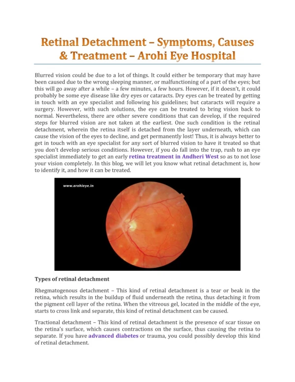
RD: • Rhegmatogenous – Greek thegma = rent • Tractional • Exudative
RD Epidemiology: • Incidence 1: 10,000 / year. • In aphakics: 1– 3%. • In the second eye (-): 5%. • In the second eye (+): 10%. • 99% of untreated symptomatic RD blindness. • 5 – 15% of population with retinal break(s) 7% of these develop new break(s).
Rhegma. RD: • Pre-requisite:- • Some degree of vitreous liquifaction. • Retinal break: - tear - hole - dialysis • Eye movements (Edie’s currents) • PVD, V-R traction
PVD_______________________________ • Due to loss of hyaluronic acid collapse of vit. collagen with liquefaction. • Rare before 30 yrs. • Increases with age (63% in > 70 yrs.) • Most PVDs are asymptomatic. 2nd eye in 1 yr. • 15% of acute PVD have a retinal tear (pathologic). • Increases significantly after cataract extraction: pathologic vs. physiologic PVD.
RD • PVD • 13-19% of PVD have vit. Hem. • PVD + hem. 70% with tears. • PVD + no hem. 2-4% with tears.
RD F/U: • Acute PVD:- • Examine periphery. • + vit. Hem. - rest, patching examine. • U/S.
RD Risk Factors: Lattice and other peripheral deg.: • Present in 8% of the population. In SA – 9.1% • As a cause of RD in 20-30%. • In RDs with L.D.:- 30-45% Atrophic holes. 55-70% A tear at edge of L.D.
RD Risk Factors: cont. High myopia: • > 6 D. • 60 yr. myope risk of RD is 2.4% whereas normal risk 0.06%
RD Risk Factors: cont. Cataract surgery: • Increases PVD: Does it convert physiological PVD to a pathological one? • 1.3% RD in aphakes. • ICCE > ECCE. • Risk of RD increased with:- - P.C. otomy: 1.3%. - Vit. loss. • 50% of RDs in 1st year.
RD Risk Factors: cont. Glaucoma: • In general population – 1% COAG. • In RD patients – 4-7% COAG. • > in pigment dispersion synd. ? myopia. • Miotics & RD.
RD Risk Factors: cont. RD in fellow eye or F/H of RD. Trauma.
RD Symptoms: • PVD – flaches & floaters. • Painless loss of peripheral VF. • Painless loss of central vision.
RD Examination: • VA. • IOP. • SLE – blood, pigment (Shafer’s sign) in the vitreous. • Careful binocular indirect ophthalmoscopy with scleral indentation. • C.L. exam.
RD Types of Breaks: • Fresh (acute) tear either:- Symptomatic tear. Tear with retinal hem. at the edge. A new tear in that location. • Flap (horseshoe) tear. • An operculated hole. • Atrophic holes.
RD DDX: • Exudative RD:- • Neoplasms. • Inflammations – VKH, ICSC, post. Scleritis. • Cong. Anomalies – optic pit, morning glory, choroidalcoloboma, nanophthalmos, uveal effusion syndrome
RD DDX: cont. • Traction RD. • Retinoschisis – senile, juvenile. • Choroidal detachment.
RD Treatment: • Prophylactic Rx:- • Olny selected breaks require Rx. • A symptomatic tear – caused by PVD or vit. Traction in the eye of a pt. C/O photopsias +/- floaters.
Indications for Prophylactic Treatment of Retinal Tears and Holes in Symptomatic Patients: Flap tears Frequently (always) Operculated holes Sometimes Atrophic holes No Macular holes Rarely Lesion Treatment _____________________________________
RD Treatment: • Prophylactic Rx to breaks:- • Cryotherapy. • Photocoagulation. • Surround it ant. & post. • Macular pucker. • Tears at margins of Rx scar.
RD Treatment: cont. • Principles of Surgery:- • Emergency. • Localization of break(s). • Creation of C-R adhestion around the break(s). • Closure of break(s). • Relief of V-R traction.