Download
1 / 22
220 likes | 467 Vues
Pathophysiology of immunity. Prof. J. Hanacek, MD, PhD. The immune system (IS). Main physiologic role: - primary role of IS is to discriminate self from nonself and to eliminate the foreign substance - finely tuned network that protects the host

E N D
Pathophysiology of immunity Prof. J. Hanacek, MD, PhD
The immune system (IS) Main physiologic role: - primary role of IS is to discriminate self from nonself and to eliminate the foreign substance - finely tuned network that protects the host against forein antigens, particularly infection agents Pathophysiologic changes of immune system: - the mentioned network can be broken down, causing IS to react inappropriatelly
Main forms of inappropriate reactions of immune system Hypersensitivity 1) Exaggerated activity against environmental antigens (allergy) 2) Misdirected activity against host’s own cells (autoimmunity) 3) Activity directed against benefitial foreign tissues, e.g. transfusion, transplants (isoimunity) Hyposensitivity 1) Activity insufficientfor protection of the body (immune deficiency)
Types of hypersensitivity • – are differentiated by the sorce of the antigens • against which the hypersensitivity is directed • Allergy – it has two facets: • a) immune response which is benefitial • b) hypersensitivity which is harmful • Definition:Deleterious effects of hypersensitive • reactions to environmental (aerogenous) • antigens expressed by disease B) Autoimmunity – disturbance in the immunologic tolerance of self-antigens – immune system reacts agaqinst self – antigens by creating autoantibodies - autoimmune diseases
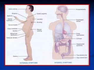
Autoimmune disease With main manifestation in: Endocrine system – hyperthyroidism (Grave’s disease) – primary myxedema (hypothyroidism) – diabetes mellitus-type 1 – Addison disease – male and female infertility – idiopathic hypoparathyroidism – partial pituitary deficiency Skin – pemphigus vulgaris – vitiligo – dermatitis herpetiformis
Neuromuscular tissues – dermatomyositis – multiple sclerosis – myasthenia gravis – postvaccinal or postinfection encephalitis – polyneuritis – rheumatic fever (heart effects) – cardiomyopathy Gastrointestinal system – celiac disease (gluten-sensitive enteropathy) – ulcerative colitis – Crohn’s disease – atrophic gastritis – primary biliary cirhosis – antibodies against intrinsic factor
Connective tissue – ankylosing spondylitis – rheumatic arthritis – systemic lupus erythematosus – polyarteritis nodosa (necrotising vasculitis) – scleroderma (progressive systemic sclerosis) Eye – Sjögren’s syndrome – uveitis Kidney – immune – complex glomerulonephritis – Goodpasture’s syndrome (basement membrane of glomerulus)
Hematopoietic system – idiopathic neutropenia, lymphopenia – autoimmune haemolytic anemia – autoimmune thrombocytopenic purpura – pernicious anemia Respiratory system – Goodpasture’s disease (interalveolar septas are influenced) • Autoantibodies are also producedby healthy individuals, particularly by the elderly. This is one of the mechanisms responsible for the ageing process (due to a deterioration of tolerance to self-antigens) • Yonger healthy individuals may produce autoantibodies without the development of overt autoimmune disease (reaction is weak)
Isoimmune disease – immune system of one individual produces an immune reaction against tissues of another individual, e.g. against transfused Er, grafted tissue, fetus during its intrauterine life Pathogenesis of hypersensitivity – it is not completly understood Main pathogenetic factors – geneticdisorders – infections – another environmental factors- polutants in air, soil, water, psychogenic stressors....
Most diseases related to hypersensitivity evolve because of interaction of at least 3 variables: a) an original insult which alters immunologic homeostasis b) the individual’s genetic makeup which determins susceptibility to the effects of the insult c) immunologic process that amplyfies the insult
Mechanisms involved in development different types of hypersensitivity Type I: IgE – mediated allergic reactions Type II: Tissue specific reactions Type III: Immune-complexes mediated reactions Type IV: Cell-mediated reactions Time corse of hypersensitivity reactions Immediate hypersensitivity reactions–within minutes Delayed hypersensitivity reactions – withinsevral hours and days from the time of exposure to antigen
Type I hypersensitivity - IgE – mediated 1) Anaphylaxis – rapid and severe reaction developed within minutes a) systemic (generalised): itching, erithema, womiting, abdominal cramps, diarhea, breathing difficulties, laryngeal edema, angioedema, vascular collapse, shock, death b) cutaneous (localised): signes of local inflammation
2) Allegy - IgE – mediated reactions Characteristics: - production of antigen – specific IgE after exposure to antigen - the most common alleregic reactions are mediated by IgE - antigens which cause allergic reactions are called allergens 3) Atopy Characteristics: - it expresses the proneness to allergy - the atopic persons produce more than normal IgE and have more Fc receptors on their mast cells - subtle defect in T-Ly function (e.g. deficiency in IgE-specific supressor cells) may account for hightened IgE production
Type II hypersensitivity - Tissue specific reactions • Characteristics: • - destruction of target cells through the action of antibodies • against an antigen on the surface of cell membrane • Explanation: • in addition to HLA system most tissue have tissue specific • antigens (TSA) = expressed only on the plasma membrane • of certain type of cells • because of limited distribution of TSA, type II disease • are limited to those tissue and organs that expresse • the particular antigen
Mechanisms involved in cells destruction in type II hypersensitivity 1) – Antibody (Atb) is bind to TSA – Atb „fixes“ complement initiation of complement cascade (CCD) lysis of the cell - e.g. autoimmune hemolytic anemia, transfusion reaction to donor blood cells 2) – Atb is bind to TSA – macrophages are able to recognize and bind the opsonised cells phagocytosis lysis of cells
3) – Atb is bind to TSA – Fc receptors on cytotoxic cells are able to recognize the antigen on the target cells binding of cytotoxic cells on target cells cytotoxic cells release of toxic substances lysis of target cells 4) – Atb is bind to TSA – Atb occupy and alters receptors on target cells blockade of normal ligands for these receptors changes in cellular functions - e.g. Grave’s disease
Type III hypersensitivity • Characteristics: • antigen-antibodies complexes (ANt-ATb-C) are created • in circulating blood deposition of ANt-Atb-C in the vessel wall • or in other extracellular tissues • this reaction is not organ – specific • harmful effect of ANt-Atb-C is caused by activation of • complement and by attempt of NE-Le to ingest these • complexes releasing of lysosomal enzymes tissue damage
Diseases caused by type III hypersensitivity • Serum sickness (called according the foreign serum used and symptoms and signs development) - general deposition of immune-complexes in blood vessels, joints, kydney - symptoms and signs: fever, enlarged lymphonodes, rash, pain • Rayanaud’s phenomenon: - temperature-dependent deposition of immune complexes in peripheral vessel (cryoglobulins) •Arthus phenomenon: - example of localised immune-complexes-mediated inflammatory response. It developes due to repeated local exposure to exogenous antigen which reacts with preformed antibodies in the vessel wall
Type IV hypersensitivity • Characteristics: • it is mediated by specifically sensitised T-Ly • it does not involve antibodies • types of sensitised Ly ivolved in reaction: • - cytotoxic T-Ly • - lymphokine-producing T-cells • Pathologic processes induced • by type IV hypersensitivity • - graft rejection - tumor rejection • - tuberculin reaction - reaction to contact with • e.g. metals or ivy
Diseases caused by type IV hypersensitivity •Rheumatoid arthritis - antigenis type II collagen in joint tissue •Hashimoto’s disease - antigen is protein present in thyroid cells •Diabetes mellitus-type 1- antigen is a protein of the -cells of Langerhans islets
Pathogenesis of hyposensitivity This disorder results from deficiciences in immunity and leads to development of different clinical manifestations. The manifestations are the result of impaired function of one or more components of the immune system – e.g. B-cells,T-cells, phagocytic cells, complement Classification: 1) Congenital (primary) immune deficiency - due to genetic disorders 2) Acquired (secondary) immune deficiency - due to another illness-e.g. cancer, viral infection - due to physiologic changes-e.g. ageing - intens stress
Diseases caused by immune deficiency • Primary T-cell defects - severe combined immune deficiency (SCID) - Di George syndrome(thymic aplasia or hypoplasia) • Primary B-cells defects - agammaglobulinemia - selective IgA, IgM, IgE deficiencies • Phagocytic defects a) quantitative defects-e.g. congenital splenic aplasia, Sickle cell anemia, congenital neutropenia b) chemotactic defects – lazy leucocyte sy. c) microbicidal defect– chronic granulomatous disease – myeloperoxidase deficiency • Complement defects