Download
1 / 22
220 likes | 239 Vues
Learn about the different types of strokes, advantages and disadvantages of CT and MRI imaging, and the general protocols for diagnosing and treating strokes.

E N D
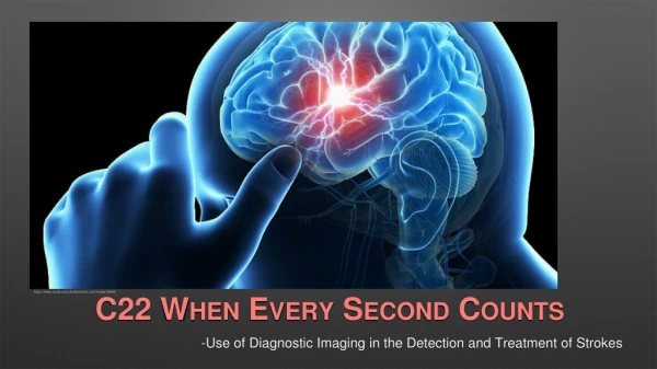
C22 WHEN EVERY SECOND COUNTS -Use of Diagnostic Imaging in the Detection and Treatment of Strokes https://www.cardiovascularbusiness.com/media/23695
OBJECTIVES • Know the 3 main types of strokes. • Know the benefits and disadvantages in utilizing CT and MRI for diagnosis. • Know the general protocol for an incoming stroke. • Know the procedures to treat the 2 main types stroke in IR.
TYPES OF STROKES TRANSIENT ISCHEMIC ATTACK ISCHEMIC STROKE HEMORRHAGIC STROKE American Heart Association https://www.strokeassociation.org/STROKEORG/AboutStroke/TypesofStroke/Types-of-Stroke_UCM_308531_SubHomePage.jsp
Approximately 85% of Strokes. Occurs when a blood vessel in the brain becomes obstructed by either a clot or fatty deposit. 2 Types: Cerebral Thrombosis Clot that forms in the brain at the area of narrowing. Cerebral Embolism Clots that forms in other parts of the body, break loose, and travel to the brain. ISCHEMICSTROKE
Approximately 13% of Strokes. Occurs when a blood vessel is weakened and ruptures, allowing blood to escape. Intracerebral - Hemorrhage within the brain. Subarachnoid - Hemorrhage between the brain and the surrounding arachnoid membrane. Can be caused by trauma, the rupture of an aneurysm, or the rupture of a malformed bundle of blood vessels. HEMORRHAGIC STROKE
Also known as a “Mini Stroke” or TIA. An Ischemic Stroke where the clot is broken up or dissolved very quickly by natural bodily processes. While only lasting a short time, TIAs can still damage brain tissue and are an indication that a more severe stroke may occur. Approximately 1/3 of people who have a TIA experience another stroke within a year. TRANSIENT ISCHEMIC ATTACKS
DIAGNOSIS OF STROKES • Once a patient has been brought into the hospital with a suspected stroke, the next step is to make a positive diagnosis to confirm a stroke and determine what type. • CT or MRI can both be used to provide imaging that will provide the doctors with the correct information. • Each modality has benefits and disadvantages when facing this type of exam protocol when time is of the essence. http://www.neuroradiologycases.com/2012/09/ischemic-stroke-and-vascular.html
CT Benefits MRI Benefits CT VS MRI • Faster acquisition ofimages than MRI. • Quicker turn around to get other patients out and prep the room. • Patients with pacemakers, pumps, or metal implants are not at risk. • Can better visualize soft tissue of the brain and vessels. • Because multiple scans are needed, MRI will save the patient a significant dose of radiation.
CT Disadvantages MRI Disadvantages CT VS MRI • Better suited to visualize bony structures and contrast than soft tissue such as the brain and blood vessels without contrast. • Higher dose of radiation with multiple scans. • If the patient is having a stroke, they might not be able give an accurate history of having a pacemaker or metal implant. • Depending on the severity of the patient’s condition, equipment being used to monitor vitals might not be MRI complatible. • If there is a patient currently in the MRI, the machine might still have 30-45 minutes of scan time left.
CT CT VS MRI WHICH WOULD YOU CHOOSE? WHICH WOULD YOU CHOOSE? MRI
CT AND MRI • With recent advancements in technology for both modalities, CT is able to image soft tissue better and MRI is able to scan faster. • Both are utilized to great effect. • Where one has a deficit, the other has an advantage. • Knowing what your hospital uses and how you can help to keep the process efficient will minimize time lost when it counts the most.
CT AND MRI • In reality, most hospitals use CT since the ability to down time the room and not worry about having a complete history of the patient helps keep the stroke protocols moving with little or no delay. With that understanding, what follows will be focused on CT and the protocols used in diagnosis. https://www.webmd.com/stroke/default.htm
When attempting to image a stroke, there are 3 exams that can be run in quick succession to answer 3 main questions. • Non-enhanced CT • CT Angiography • Perfusion CT • Ischemic Stroke or Hemorrhagic Stroke? • Is flow to a major vessel blocked? • What tissue can be saved and what is already damaged? IMAGING PROTOCOLS
Un-enhanced is a CT done without the use of contrast. Essentially, it is a 3D X-ray and shows overall anatomy in the area of interest. A Perfusion CT shows what areas of the brain are being supplied with blood and which areas are not. This is done with a program that recognizes an increase or decrease in blood flow rate. A CT Angiogram is done with intravenous contrast injected and timed specifically to highlight the blood vessels in the area of interest, in this case, the brain. IMAGING PROTOCOLS https://www.ispn.guide/wp-content/uploads/2017/12/5.CT-angio-coronal.jpg https://radiopaedia.org/images/889537 http://file.scirp.org/Html/5-1780107_41505.htm
TREATMENT Once the imaging is completed and a stroke is positively diagnosed, as well as what type, options for treatment become available. In the case of an Ischemic Stroke, medications known as Thrombolytics will be given to help try and break up the clot. In many cases, surgery is necessary to either clear the clot or repair the ruptured blood vessel. The patient will then be moved to Interventional Radiology for the procedure.
TREATMENT • In many hospitals, the procedure will take place in a surgical suite with a biplane digital x-ray machine. This allows for 2 images to be taken at the same time from opposing views. The ability to pulse the x-rays giving a live image allows for real-time feed back for the doctor. • Whether the stroke is Ischemic or Hemorrhagic, this will help the doctor and IR technologists guide tools such as balloons and stents through arteries and vessels to reach the problematic area. https://www.shimadzu-medical.eu/trinias-b8-mix-package
TREATMENT Depending on the type of stroke, one of two procedures will take place under x-ray guidance. Ischemic Stroke • For occluded vessels, a process known as Angioplasty will be used to guide a catheter to the clot. A thrombolytic will be delivered directly and a small balloon on the end of the catheter will be inflated to once again allow blood flow and widen the artery. The placement of a stent will also ensure the vessel will stay open. https://www.youtube.com/watch?v=Ew9VlBBBa0Y
TREATMENT • Hemorrhagic Stroke • If lucky, the scan is able to find weakened vessels before they rupture. When that happens, a procedure known as an Endovascular Coil Treatment will be done. This involves guiding a catheter with the to the site of the aneurysm and feeding a length of wire into the area, letting it coil, and helping begin the clotting process. A stent may also be placed if the aneurysm is larger in size to contain the coils.
TREATMENT • Hemorrhagic Stroke • Once a vessel has ruptured, immediate action needs to be taken to stop the bleeding and relieve any pressure on the brain. A surgical clip will be placed at the site of the hemorrhage and any excess blood in the area will be removed at the time of surgery, or over time with a drain. • This procedure is done by a neurosurgeon, and not usually assisted with the use of X-ray. https://www.kaleidahealth.org/services/display.asp?pt=G&p=1357&s=695
Conclusion • With the advent of fast and reliable imaging, stroke diagnosis times have been drastically reduced. This in turn has increased the survivability rate of strokes that would have otherwise ended in death. • Interventional Radiology procedures have also benefited from the use of x-ray guidance to reduce patient time on the table as well as keeping these operations minimally invasive to reduce rehab timelines.
Know the 3 main types of strokes. Ischemic, Hemorrhagic, and Transient Ischemic Know the benefits and disadvantages in utilizing CT and MRI for diagnosis. Both have qualities necessary to image correctly, but CT is the current front runner for diagnosis Know the general protocol for an incoming stroke. Unenhanced CT, Ct Angiogram, and Perfusion CT Know the procedures to treat the 2 main types stroke in IR. Ischemic Stroke = Angioplasty Hemorrhagic Stroke = Endovascular Coil Treatment or Clipping Overview
References Enrique Marco de Lucas, Elena Sánchez, Agustín Gutiérrez, Andrés González Mandly, Eva Ruiz, Alejandro Fernández Flórez, Javier Izquierdo, Javier Arnáiz, Tatiana Piedra, Natalia Valle, Itziar Bañales, Fernando Quintana. CT Protocol for Acute Stroke: Tips and Tricks for General Radiologists. RadioGraphics. 2008; 28:1673-1687. doi: 10.1148/rg.286085502 Tatlisumak T. Is CT or MRI the Method of Choice for Imaging Patients With Acute Stroke? AHA Journals. Stroke. 2002;33:2144-2145. doi: 10.1161/01STR.0000026862.4244.AA Mayo Clinic. Stroke Diagnosis and Treatment. https://www.mayoclinic.org/diseases-conditions/stroke/diagnosis-treatment/drc-20350119. Accessed October 2, 2018. National Stroke Association. Hemorrhagic Stroke Fact Sheet. http://www.stroke.org/stroke-resources/library/hemorrhagic-stroke. Accessed October 2, 2018. American Stroke Association. About Stroke. Types of Strokes. https://www.strokeassociation.org/STROKEORG/AboutStroke/TypesofStroke/Types-of-Stroke_UCM_308531_SubHomePage.jsp. Accessed October 13, 2018. Society of Interventional Radiology. Stroke. https://www.sirweb.org/patients/stroke/. Accessed October 13, 2018. RadiologyInfo.Org. Stroke. https://www.radiologyinfo.org/en/info.cfm?pg=stroke. Accessed October 11, 2018.