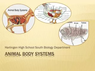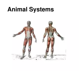
Animal Systems
Explore the essential components of animal biology, focusing on various tissue types and organ systems. This guide covers epithelial, muscle, connective, and nerve tissue types; the structure and function of organs including the stomach and heart; and the interconnected nature of organ systems such as the digestive and circulatory systems. Learn how these tissues and organs work collaboratively to fulfill vital functions in the body, including digestion and blood circulation, and the significance of maintaining overall health.

Animal Systems
E N D
Presentation Transcript
Tissues • A collection of similar cells that perform a particular function.
Epithelial tissue • Thin sheet of tightly packed cells that covers the body surfaces and lines internal organs • Skin • Digestive system lining • Muscle tissue • Specialized tissues that contain proteins and can contract and enable the body to move • Muscles that make bones move • Muscles in the digestive tract • Heart muscles • Connective tissue • Specialized tissue that provides support and protection for various parts of the body • Bone • Tendon • Blood • Nerve tissue • Specialized tissue that conducts electrical signals from one part of the body to another • Brain • Nerves
Organs • An organ is a structure composed of different tissues working together to perform a complex body function. • Stomach • Heart • Eyes • Skin
Organ Systems • A system of one or more organs and structures that work together to perform a major vital body function. • Digestive system • Reproductive system • Muscular/skeletal system • Respiratory system
Alimentary canal • Mastication-the act of chewing (breaking down food into smaller pieces) • Saliva moistens food but also contains the enzymes amylase and ptyalin to break down carbohydrates starchy foods
Food leaves the mouth and is pushed down the esophagus by the tongue. • Pharynx- contains opening to trachea as well. Epiglottis covers opening of trachea. • Esophagus-muscular tube dorsal to trachea • Relies on a rhythmic wave-like motion called peristalsis
Food enters the stomach from the esophagus by passing through the cardiac sphincter. • The stomach lining has glands that secrete gastric juices (HCl to kill bacteria and Pepsin to break down complex protiens).
Stomach is protected by a layer of mucus. • Food and juices mixture is called chyme and sits in stomach for 1-4 hours.
Small Intestine • The majority of chemical digestions occurs here as various juices are added to change the food.
Bile is made by the liver and stored in the gall bladder. The bile duct carries the bile from the gall bladder to the small intestine. Bile emulsifies fats into small droplets. • Pancreatic juice is made in the pancreas and added to the small intestine through the bile duct. Pancreatic juice neutralizes stomach acid and contains enzymes that carry out digestion • Intestinal juice is made in the lining of the small intestine and contains enzymes that break down sugars (maltase breaks down grain, lactase breaks down dairy, and fructase breaks down fruit).
Digestion is complete in the first section of the small intestine, called the duodenum. Usable materials are picked up by the blood throughout the rest of the small intestine. • Unusable products are moved by peristalsis to the large intestine.
Large Intestine • The large intestine is shorter than the small intestine but is large in diameter. • The appendix is found where the small and large intestine join. • The large intestine absorbs water from the undigested food. • Diarrhea is when peristalsis is too fast and not enough water is absorbed. • Constipation is peristalsis is too slow and too much water is absorbed.
Rectum and Anus • The rectum is the muscular section found at the end of the large intestine. The rectum pushes solid waste (faces) through the anus.
Diseases • Appendicitis • Cholecystitis- gall bladder • Cirrhosis- destruction of liver cells • Diarrhea • Diverticulitis • Gasteroenteritis • Hernia • Pancreatitis • Liver transplant • Ulcer • Colitis • Bariatric surgery
Salivary Gland Teeth Tongue Epiglottis Esophagus Liver Stomach Gall Bladder Bile Duct Duodenum Pancreas Transverse Colon Ascending Colon Small Intestine Descending colon Appendix Rectum
Circulatory System - Blood • Blood is the fluid that supplies essential substances to all body cells and removes waste products. Blood is made of four parts • Plasma (about 90% water) • Red blood cells (numerous, contain hemoglobin, which combines with oxygen to give a rich red colour) • White blood cells (destroy pathogens) • Platelets (help prevent blood loss from injuries)
Blood flows through three main vessels: • Arteries – carry blood away from the heart • Veins – carry blood to the heart • Capillaries – thin walled vessels that allow oxygen, food and water to be exchanged with the blood. • Blood pressure is the force exerted by the flowing blood against the walls of the vessels.
Blood is continually circulated throughout the body. • Cardiac circulation is oxygen rich blood that feed the cardiac muscles • Pulmonary circulation is blood that leaves the heart and goes directly to the lungs. • Systematic circulation is the blood that leaves the heart from all points of the body other than heart and lungs
Circulatory System - Heart • The heart is a muscular pump that beats continuously and rhythmically. • The heart rate is the number of times the heart beats per minute. • The lubb-dubb sounds are caused by the opening and closing of the heart valves. • Changes in breathing correspond to changes in heart rate
The heart is surrounded by a protective sac called the pericardium. • The heart has four chambers – two upper chambers are the atria and two lower chambers are the ventricals. The left side is separated from the right side by the septum.
Blood travels from the left ventricle to all body cells via the aorta, the main artery of the body. • The blood that leaves the left ventricle is rich in oxygen and provides the needed oxygen for all body systems (bright red) • Some of this blood travels back to the heart as described in cardiac circulation,
The blood from the body eventually returns to the right atrium of the heart through large veins – the superior vena cava and the inferior vena cava (systematic circulation) • Blood that circulates from the right ventricle of the heart through the pulmonary artery to the lungs and back to the left atrium is involved in pulmonary circulation.
When blood returns to either atria, it enters the chamber and remains until pumped through the ventricle. • From the right atrium to the right ventricle, the blood is pumped through the tricuspid valve. • Blood from the left atrium to the left ventricle is pumped through the bicuspid valve
Disorders of the Circulatory System • High blood pressure • Heart attack and angina pectoris • Stroke • Arteriosclerosis • Rheumatic fever • Anemia • Leukemia
Respiratory System Air is a mixture of Nitrogen (78%), Oxygen (21%), water vapour (1%) and a few trace amounts of other gases. Every minute you breath in about 6L of air. Cells need air to survive. They need the oxygen to burn energy. The energy releasing process that is fuelled by oxygen is called respiration.
The respiratory system is responsible for getting oxygen into the body and removing carbon dioxide from the body.
Air contains dirt, bacteria and pollutants. • It is healthier to breath through your nose because it warms the air and the mucus filters dust and microorganisms. • Air travels through the nose then the trachea and through the bronchi in a few short seconds. These lead to the lungs. • The epiglottis covers the path to the digestive system while opening the passage to the respiratory system.
The trachea is a bumpy tube-like structure. The bumps are rings of cartilage and the tube is lined with mucus to catch dust and bacteria. Small finger-like cilia return this dusty mucus to the mouth or nose fro removal. • At the top of the trachea is a box structure called the larynx. It too is made of cartilage. Men usually have larger voice boxes which produce a lower sound.
Inside the larynx are the vocal chords, which are small folds of tissue that stretch. They vibrate to produce your voice. • A high voice is due to tight chords that vibrate quickly. A low voice is due to loose chords that vibrate slowly.
From the trachea, air enters the left and right bronchi and then into each lung. • Branches of the bronchus break into bronchioles which have alveoli on the end, small sacs where oxygen and carbon dioxide are exchanged.
Muscles attached to your ribs contract and lift the ribcage up and out. At the same time, the diaphragm contracts and flattens. This causes your chest to expand. • The diaphragm is a dome shaped muscle that contracts involuntarily. • Nerves that control the diaphragm can become irritated (eating to fast, laughing to hard). As you inhale, the space between the vocal chords snap shut with a clicking sound (a hiccup)