Body Cavity fluids
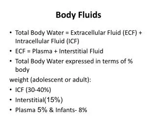
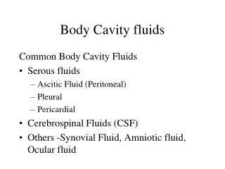
Body Cavity fluids. Common Body Cavity Fluids Serous fluids Ascitic Fluid (Peritoneal) Pleural Pericardial Cerebrospinal Fluids (CSF) Others -Synovial Fluid, Amniotic fluid, Ocular fluid. Sample Collection. CSF – Lumbar puncture- 3-4 tubes in plain sterile tubes

Body Cavity fluids
E N D
Presentation Transcript
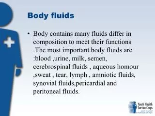
Body Cavity fluids Common Body Cavity Fluids • Serous fluids • Ascitic Fluid (Peritoneal) • Pleural • Pericardial • Cerebrospinal Fluids (CSF) • Others -Synovial Fluid, Amniotic fluid, Ocular fluid
Sample Collection • CSF – Lumbar puncture- 3-4 tubes in plain sterile tubes • Serous fluids – EDTA for cell counts and morphology for other tests in Heparin or blood culture tubes
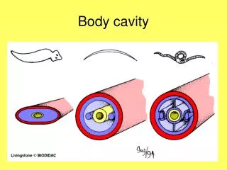
PLEURAL / ASCITIC FLUID Pleural cavity normally contains small amounts of fluid facilitating movement of parietal & visceral pleura, which are lined by mesothelium. Peritoneum is lined by mesothelium and contains small amounts of peritoneal fluid. Increased accumulation of fluid is called as Ascites.
GROSS EXAMINATION • Quantity • Colour • Appearance • Clot • Turbid fluids - Supernatant Clear - Cellular elements • Hazy -Chylous (obst. of Thoracic duct)
MICROSCOPIC EXAMINATION Cell counts, if necessary, with dilution Differential counts Normal cells of Pleural Fluids • Macrophages • Mesothelial cells • Lymphocytes • Monocytes
MESOTHELIAL CELLS • Bland cells forming a monolayer covering serous surfaces of body cavities. • 20 - 40 microns in diameter • Round to oval nuclei, inconspicuous nucleoli, cytoplasm exhibits varying degrees of peripheral vacuolization, ‘Feathery appearance’. Two cells joined by ‘window’. • Irritated by inflammation, chemical agents & trauma. Cells enlarged with nuclear atypia.
CEREBROSPINAL FLUID (csf) • Collection of specimen: 3 tubes • Cell count, Cytomorphology, Cytochemistry • Biochemistry • Microbiology Specimen should be processed within one hour of sample collection
Material required: Procedure: • WBC DILUTING FLUID(Turk’s Fluid): • Methylene Blue(30mg/ml) • Glacial acetic acid • Distilled water Neubauer chamber:
Calculation of Cell count N X Dilution factor Total cell count = Area of total squares counted X Depth Correlation of cell count with cytomorphological findings is essential. QUALITY CONTROL IN OUR LAB
CEREBROSPINAL FLUID • Derived from filtration and secretion through choroid plexus, produced at the rate of 500 ml/day. • Collects wastes, circulates nutrients and lubricates CNS. • Normal CSF volumes: • In Adults: 90 - 150 ml • In Neonates: 10 - 60 ml • Normal Leukocyte counts: • In Adults: 0 - 5 cells/cumm • In Neonates: 0 - 30 cells/cumm INDICATIONS FOR LUMBAR PUNCTURE • Infections • Malignancy • SAH • Demyelinating diseases
GROSS EXAMINATION • Quantity • Colour • Appearance • Clot formation • Coagulum • Xanthochromia
MICROSCOPIC EXAMINATION PREPARATION BY CYTOCENTRIFUGE Other techniques Sedimentation Cell catch Filtration
PARTS OF CYTOSPIN • Auto-locking, plastic outer lid • Autoclavable Sealed Head • Disposable sample chambers with caps • Safety alarms that protect users and specimens • Wipe-clean control panel
PRINCIPLES OF CYTOSPIN Cytocentrifuge is a microprocessor controlled cell preparation system that uses centrifugal forces to deposit cells onto the slide
Using centrifugal principles, the Cytospin deposits cells onto a clearly-defined area of a glass slide and allows for the absorption of the residual fluid into the sample chamber’s filter card. • Cytocentrifugation also constructively flattens cells for excellent nuclear presentation. • During operation, the instrument’s spinning action tilts Cytofunnels upright and centrifuges cells onto the deposition area of the slide, giving all cell types equal opportunity for presentation.
Load up to 200 µl of this suspension in each cuvette. • Spin at 800 rpm for 3 min ( 500 rpm/ 4 min) • Extract the slide, paper and cuvette without disarranging. • Carefully detach the cuvette and the paper without damaging the fresh cytospin. Hold firmly together glass slide and cuvette when extracting from metal holder. • Mark the area around the cytocentrifuged cells with dry point or permanent marker. • Proceed with either immediate fixation or drying. Store unfixed cytospins for max 2 days at room temperature.
Normal cells of CSF • Lymphocytes and monocytes are normally present in small numbers in a ratio of 70:30. Monocytes are more in number in neonates and children. • Choroid plexus and ependymal cells are rarely seen in hydrocephalus and after intra-thecal chemotherapy • Cartilage, ganglion cells and artificial admixture of hematopoietic cells. • Contaminants : fungus and bacteria.