Download
1 / 1
10 likes | 131 Vues
Phase association and binding energetics of SWCNTs into phospholipid Langmuir monolayers. Peter N. Yaron 1 , Philip A. Short 2 , Brian D. Holt 2 , Goh Haw-Zan 3 , Mohammad F. Islam 1,4 , Mathias Lösche 2,3 , Kris Noel Dahl 1,2

E N D
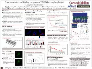
Phase association and binding energetics of SWCNTs into phospholipid Langmuir monolayers Peter N. Yaron1, Philip A. Short2, Brian D. Holt2, Goh Haw-Zan3, Mohammad F. Islam1,4, Mathias Lösche2,3, Kris Noel Dahl1,2 1Chemical Engineering, 2Biomedical Engineering, 3Physics, 4Materials Science and Engineering, Carnegie Mellon University, Pittsburgh, PA Introduction Langmuir Monolayers Fluorescence Lifetime Imaging Microscopy (FLIM) Tethered Bilayer Membrane (tBLM) • Single-walled Carbon nanotubes (SWCNTs) have been identified as promising candidates for targeted drug delivery due to their low toxicity and ability to be functionalized using various bioactive groups • Currently undetermined what mechanical and biological mechanism(s) are responsible for uptake into cells • Objective: Determine the predominant membrane insertion and cellular uptake mechanism of SWCNTs • Lipid phase behavior can be controlled changing surface area, A, affecting surface pressure, P Image courtesy of H. Nanda NCNR NIST 0000 ≤ tm ≤ 1000 ps • Fluorescence emission lifetime is a characteristic of every fluorophore • Lifetime also sensitive to the nanoenvironment: pH, [O2], binding to macromolecules, etc. • HeLa cells transfected with pAcGFP1-Endo • Incubated with SWCNTs at 100 µg/ml for various time points • Changes in fluorescence lifetimes were observed in SWCNT-treated cells 1000 ≤ tm≤ 2000 ps 2000 ≤ tm≤ 3000 ps Isotherm and Phase Diagram of DPPC monolayer liquid condensed, LC liquid expanded, La Endosome count after SWCNT incubation Equivalent Circuit two-dimensional gas, LG Error bars are the standard deviation from the average values of the data sets tBLM capacitance n = 32 n = 33 = SWCNT synthesis spreading resistance • Synthesized by HiPCO (high-pressure carbon monoxide conversion synthesis) • Size selected using density gradient length sorting • Highly purified sorting to remove carbonaceous polymorphs and metallic catalyst particles • Stabilized and dispersed using a biocompatible tri-block co-polymer Pluronic F127 n = 18 n = 17 endosomes/cell EIS Spectra = n = 33 n = 30 tBLM resistance substrate interfacial impedance n = 35 stray capacitance 16:0 PC (DPPC) A) 25 20 15 control 10 5 0 Maximum Insertion Pressure (MIP) time after treatment (min) C) • Measuring the change in surface pressure after exposure to SWCNTs from different starting pressures one can extrapolate the maximum insertion energy needed for a SWCNT to penetrate a phospholipid monolayer Bode plots (A & B) of tBLMs with SWCNTs (red) and without (black), (C) Cole-Cole plot (C) of the tBLM after incubation with SWCNTs B) SWCNT Dimensions Conclusions radius : 0.7 – 1.3 nm • Fixed cell imaging shows an increase in the number of endocytotic vessels • FLIM shows altered lifetime of GFP labeled endosomes suggesting SWCNT uptake via endocytosis • Langmuir monolayers yield a maximum insertion pressure of 28 mN/m which is below MIP needed for BLM insertion (~30 mN/m) • EIS shows negligible changes in capacitance and resistance indicating minimal incorporation of SWCNTs by purely physical mechanisms Solvent Maximum Insertion Pressure FLIM of GFP Labeled Endosomes + SWCNTs Distal leaflet Control 5 min 25 min mean length : 145 ± 17 nm Fixed Cell Imaging Proximal leaflet A SWCNTs MIP • HeLa cells were transfected with pAcGFP1-Endo and incubated with 100 mg/ml of SWCNTs (A) • Endocytotic vessels were determined by intensity maxima in the GFP fluorescence filter range using Image J (B) Aqueous Reservoir Tether Lateral Spacer Electrochemical Impedance Spectroscopy (EIS) References and Acknowledgements B • EIS was performed on tethered bilayer membranes before and after incubation with SWCNTs • changes in tBLM due to inclusion of SWCNTs can be related to changes in capacitance and resistance (A-C) [1] Holt et al. ACS Nano. 4, (2010): 4872-4878 [2] Bianco, et al. Curr. Opin. Chem. Bio.9, (2005): 674–679 [3] Kostarelos et al. Nature nano. 108, (2007): 108-113 [4] Gao, et al. Proc. Nat.Acad. Sci. 102, (2005): 9469-9474 [5] S. Pogodin et al. ACS Nano. 4, (2010): 5293–5300 Funding: NSF CAREER, NIH (1P01AG032131) Image Statistics of Fluorescence Lifetimes Biological & Biophysical Basis of Membrane Dynamics and Organization workshop, Nov. 5 & 6, Mellon Institute of Science Control 5 min. 25 min.