Download
1 / 59
590 likes | 729 Vues
Disorders of Small & Large Bowel (Specially dedicated to Mark Wahba -- hope this helps buddy). Moritz Haager April 01, 2004. Outline. Appendicitis Useful & useless tests Mesenteric Ischemia When to suspect it & how to chase it Diverticular disease Who has it? Whom to Tx & how. Case 1.

E N D
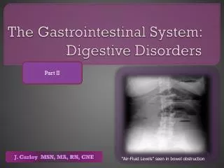
Disorders of Small & Large Bowel(Specially dedicated to Mark Wahba -- hope this helps buddy) Moritz Haager April 01, 2004
Outline • Appendicitis • Useful & useless tests • Mesenteric Ischemia • When to suspect it & how to chase it • Diverticular disease • Who has it? Whom to Tx & how.
Case 1 • 25 yo female c/o right lower quadrant pain. • What is your differential diagnosis? • How does you DDx change if she is 5 yo? 85 yo? Male? • Which historical & physical exam features are helpful in narrowing it down? • What lab & DI tests are useful? • Who can go home, who should stay, who needs to go to the OR?
Appendicitis Fast Facts • Incidence 1/1000 • Lifetime risk 6% -- genetic predisposition • Mortality 0.1% (20-60x higher in perf’d) • Initially misdiagnosed in 30% • Accepted negative laparotomy rate 20-25% • Perforation rate 20% • 70% in <9 yo or > 60 yo
True or False? • Appendicitis can occur at any age • T: most common in 10-30 yo • Appendicitis can present w/ LUQ pain • T: rare but possible (0.06%) • Appendicitis does not recur • F: estimated to recur in 6% • Definite gastroenteritis rules out appendicitis • F: viral & other infections can actually cause appendicitis due to lymphoid hypertrophy
Gastroenteritis Crohn’s Testicular torsion Meckel’s diverticulum Diverticulitis Mesenteric adenitis Cholelithiasis Pancreatitis Bowel obstruction Pelvic inflammatory disease Endometriosis Ovarian cyst Tubo-ovarian abscess Ectopic pregnancy Mittelschmerz Pyelonephritis Urinary Tract infection Appendicitis DDx
History & Physical • Aim to differentiate pts into 3 groups • High suspicion for appendicitis – need for immediate surgery • i.e. classic presentation • Intermediate suspicion for appendicitis – no clear-cut need to go to OR yet • Atypical presentation • Low suspicion for appendicitis
Diagnostic Strategies The only absolutely lab necessary test • Hx & P/E • Labs: CBC & diff, CRP, β-HCG, U/A, etc • Radiography • Plain films • Ultrasound • CT • Observation & serial examination • Laparoscopy
Classical Presentation • Good story: Pain starts as vague peri-umbillical discomfort localizes to RLQ as sharp pin-point pain • WBC > 10 • Rebound & guarding at McBurney’s Point • Associated N & V, anorexia • Fever • Present within 48 hrs of onset • Occurs in only 50-60% of patients
If you didn’t know & were to embarrassed to ask • Psoas sign • With pt supine, get pt to flex hip against resistance by pushing down against knee -- pain = +ve • Obturator sign • Passively flex hip & knee and internally rotate leg at the hip -- pain = +ve • Rosvings sign • press down in LLQ then release suddenly -- pain = + ve
Symptoms Abd pain 97-100% Anorexia 70-92% Nausea 67-78% Vomiting 49-74% RLQ migration 49-61% Fever 10-20% Diarrhea 4-16% Constipation 4-16% RLQ pain LR+ 8.0; LR- 0.2 Rigidity LR+ 4.0; LR-0.82 Migration LR+ 3.1; LR- 0.5 Previous similar pain LR- 0.3 Signs Abd tenderness 95-100% RLQ tenderness 90-95% Rebound 33-68% Rectal tenderness 30-40% Cervical motion tenderness 30% Rigidity 12% Psoas sign3-5% Obturator sign 5-8% Rosvings sign 5% Palpable mass <5% Avg Temp 37.9oC Signs & Symptoms Wagner et al. Does this patient have appendicitis? JAMA 1996; 276: 1589-94
Accuracy of clinical findings Wagner et al. Does this patient have appendicitis? JAMA 1996; 276: 1589-94 * based on only one study
Do you need to do a Rectal? • NO • Sens 41%, Spec 77% • LR+ 0.83 - 5.34 • LR- 0.36 - 1.15 • Wagner et al. Does this patient have appendicitis? JAMA 1996; 276: 1589-94 • “Pain on rectal palpation has no discriminatory or predictive power” • Andersson. Meta-analysis of the clinical and laboratory diagnosis of appendicitis. Brit J Surg. 2004; 91: 28-37
Utility of the WBC count • Elevated in 70-90% of pts w/ appendicitis • Somewhat helpful if >19 or <7 but this happens in only ~20% of pts • Snyder & Hayden. Accuracy of leukocyte count in diagnosis of acute appendicitis. Ann Emerg Med. 1999; 33: 565-574 • Very non-specific – many of the other disorders on the DDx will have elevated white count
Andersson. Meta-analysis of the clinical and laboratory diagnosis of appendicitis. Brit J Surg. 2004; 91: 28-37 • No clinical or laboratory parameter alone found to have sufficient discriminatory or predictive capacity • Performance increased considerably when 2 or more variables were combined • Most useful variables were clinical markers of peritonitis, pain migration, and WBC & diff combined w/ CRP • Caveat: highly selected population of pts w/ suspected appendicitis admitted for further evaluation
Radiography • Plain films • 0% sensitivity for appendicitis– a waste of time if appy is your 1st thought • Ultrasound • Sensitivity 75-90%, Specificity 86-100% • Able to identify alternate diagnoses esp. in female pts • CT • Sensitivity 90-100%, specificity 91-99% • Able to identify alternate diagnoses
Ultrasound (Graded Compression) • Test Characteristics • Sensitivity 75-90%, Specificity 86-100% • Pros • No radiation, safe in kids, pregnant pts • Can identify alternate Dx esp. in female pts • Cons • Difficult for us to get locally • Operator-dependant • Limited in obese pts or ++ bowel gas • Identifies alternate Dx less often than CT • Painful
CT • Test characteristics • Sensitivity 90-100%, specificity 91-99% • Pros • Identifies alternate Dx more often than U/S • Fast & accessible in our practice setting • Cons • Radiation dose (~100 CXR’s) • Multiple techniques in literature: controversial as to which is best but all ~90-100% sensitive • Spiral vs. conventional • Focused vs. entire abdomen • Unenhanced, various combinations of IV, oral, rectal contrast • Less accurate in pts w/ little intraabdominal fat
Which test is better? • 2 prospective RCT’s of U/S vs. CT • CT more sensitive & specific than U/S • 94-97% sensitive vs. 76 – 100% for U/S • 100% specificity vs. 76-90% for U/S • More alternate Dx identified by CT • Horton et al. Am J Surg 2000; 179: 379-81 • Walker et al. Am J Surg 2000; 180: 450-55
Which test is better? • 120 consecutive pts 8-81 yo w/ ?appy who were to well to go to OR but too ill to simply D/C • Did focused CT w/ rectal contrast & U/S within 1 hr on all pts • Gold standard was pathology or clinical f/u x 6 mo • CT: 95% sensitive, 89% specific • U/S: 87% sensitive, 74% specific • CT identified 14 alternate Dx vs. 9 for U/S • U/S missed 2/3 of pts w/ perforation • Pickuth et al. Suspected acute appendicitis: Is ultrasonography or computed tomography the preferred imaging technique? Eur J Surg. 2000; 166: 315-19
Does imaging change mgmt? • 2 studies of CT in pts w/ suspected appendicitis comparing Tx plan before & after access to results of scans • CT changed disposition in 27 – 59% of pts • Prevented d/c of ~3% pts w/ appendicitis • Prevented negative laparotomy in 3-13% • Alternate Dx in 11-20% • Frank et al. Unenhanced helical CT scanning of the abdomen and pelvis changes disposition of patients presenting to the emergency department with possible acute appendicitis. J Emerg Med 2002; 23: 1-7 • Rao et al. Effect of computed tomography of the appendix on treatment of patients and use of hospital resources. N Eng J Med. 1998; 338: 141-6
Bottom-line • Pts w/ high pre-test probability should go for appendectomy regardless of imaging result • Pts w/ very low pre-test probability should be clearly instructed when to return for re-evaluation • Pts who fall in b/w these extremes benefit most from imaging
Bottom-line • Both are good but if I had only one test I’d take the CT • If I had only 5 CT’s or U/S’s available per month I’d use them on women rather than men • If I had a very skinny pt, pregnant pt, or kid I’d prefer to do a U/S • If I thought the main DDx was gastro vs. appy I’d be happy w/ a focused CT …. • …but if I had the sense this pt has something going on which could be appy but I’m really not sure I’d want the entire belly CT preferably with contrast
Observation & Serial exams • Reasonable alternative but not well studied • Pro • Observation for 6-10 hrs in intermediate-risk pts does not appear to increase risk of perforation while potentially lowering negative appy rate • Con • In kids perforation is the rule – delays may increase complications • Cost of admission may outweigh cost of DI • If observed in the department can slow flow
Proposed Appy Algorithm Paulson et al. Suspected appendicitis. N Eng J Med 2003; 348: 236-42
Case 2 • 61 yo female with severe acute abdominal pain for 3 hrs. Has vomited and had 3 watery stools. • Afebrile, no evidence of peritonitis • PMHx remarkable for rheumatic fever & HTN.
Mesenteric Ischemia • Simply stated this is blocked or restricted blood flow to the gut • Pathophysiology essentially same as that for CAD & thromboembolic cardiovascular dz • 4 major types w/ different Tx & prognosis • Occlusive (80%) • Arterial • Embolic (50%) • Thrombotic (15%) • Venous • Thrombotic (15%) • Non-occlusive (20%) • Low flow states e.g. sepsis, hypovolemia “shock bowel”
Anatomy • Celiac Trunk • Pharynx, esophagus, stomach, proximal duodenum, liver, GB, pancreas, spleen • SMA • Distal duodenum, jejunum, ileum, cecum, ascending colon, 2/3 transverse colon • IMA • Distal 1/3 transverse colon, descending & sigmoid colon, rectum • Extensive collateral supply & overlap exist b/w these = protective to a large degree • Venous system parallels arterial system
Clinical Presentation • Triad of acute abdo pain, diarrhea, & vomiting in high risk pt • Very non-specific especially early when its critical to make the Dx – broad DDx • ‘Time is gut” & dead gut frequently = dead pt • Peritoneal signs = transmural necrosis • 70-90% mortality untreated • 45-50% mortality treated w/ peritonitis • ~10% mortality if early Dx (no peritonitis) • Most useful S & S: • Visceral pain out of proportion to exam • Dull, not worse w/ movement or palpation • Older & Risk factors for atherosclerotic +/- embolic dz
Mesenteric arterial embolic Dz • Vast majority involve SMA (45o angle) • Better prognosis than thrombosis • Emboli lodge at distal branch points rather than origin smaller area of gut involved • Tend to present earlier w/ more typical Sx • Better response to Tx • Risk Factors • Older, CAD, Post-MI (mural thrombi), CHF, a fib, valvular Dz, aortic dissection / aneurysm, aortic surgery, angiography, Hx of thromboembolic Dz
Mesenteric arterial thrombosis • Occurs in more proximal vessel origin worse prognosis • SMA again most common site • Analogous to CAD: angina & MI • May have Hx of abdominal angina • Strong association w/ CAD
Mesenteric venous thrombosis • Younger pts (can occur any age) • Less mortality (20 – 50%) • 95% of all cases involve SMV • Risk factors • Hypercoagulable state • Polycythemia vera, myeloproliferative Dz, ATIII deficiency, protein C & S deficiency, DVT, malignancy, estrogen Tx, pregnancy, sickle cell • Intraabdominal inflammation • Pancreatitis, diverticulitis, appendicitis, cholangitis • Trauma • Other • CHF, renal failure, the bends, portal HTN
Non-occlusive mesenteric ischemia • Due to mesenteric vasoconstriction or low-flow state secondary to other critical illness • CVS • CHF, MI, Post CABG, • Shock states • Septic, hypovolemic, cardiogenic etc • Drugs • Inotropes, cocaine, ergots, digoxin
Diagnostic Strategies • History & physical • Labs • WBC, lactate, CK • Diagnostic Imaging • Plain films • CT • U/S • Angiography
Utility of lab tests • WBC • Elevated in most but decreased sensitivity early, & very non-specific • CK • 54% sens at 2 hrs, 75% sens at 4 hrs, 83% spec • Lactate • Up to 96- 100% sensitive, 42% specific (at what time point) • ? α-Glutathione S-transferase • Promising but limited studies at the moment • Paucity of good studies on markers • Generally too insensitive & non-specific
Plain films • Only 28-30% sensitive • Many non-specific findings.. • Ileus, free air, obstruction • …or specific findings (too) late • Pneumatosis intestinalis, portal venous gas, thickened bowel wall, thumbprinting • Too insensitive & nonspecific to aid in early Dx
CT • Sens 64-82% • Look for evidence of ischemia in bowel wall & mesentary • Evidence of clot in SMA • First investigation done routinely here • If suspecting mesenteric ischemia very important to let your radiologist know • Good but not good enough • If CT is negative & high pre-test probability you need an angiogram
Ultrasound • Doppler can determine major obstruction to flow in both venous & arterial systems • See dilated, tubular vessels full of echogenic material (clot) and abnormal flow • Limitations • Used & studied primarily in venous thrombosis & chronic mesenteric ischemia • Really don’t know much about how it performs for acute mesenteric ischemia • Only good for more proximal blockages • Has the usual limitations inherent to all U/S exams
Angiography • Gold standard test (~90% sens) • Diagnostic & therapeutic • Infusions of vasodilators into SMA (papaverine) • Angioplasty • Controversies: • When & on whom to do it • Drawbacks • Time-consuming • Risks of contrast & invasive procedure • Expensive
Angiography: Early vs. late strategy • Most authors feel angiography should be done early in pts w/o peritonitis & high suspicion • Can buy time (papaverine) • Can aid in surgical decision making • Surgical: embolectomy, thrombectomy, endarterectomy, bypass grafting • Non-surgical: angioplasty • Early (before peritonitis) angiography & intervention decreased mortality from 70-90% to 10% in several studies • Down side is high rate of negative angios & associated risks & costs
Angiography: When not to do it • Contraindicated in: • Unstable hypotensive pts on vasopressors • Difficult to differentiate b/w occlusive & non-occlusive etiologies • Can’t infuse vasodilators • Pts w/ peritonitis • Delays surgery
MRI • Gadolinium-enhanced MRA appears to be very good • MRI best at differentiating potentially viable from dead gut • Currently limited by length of acquisition time & cost but will likely play larger role in the future
ED management • ABC’s • Maintain CO • Maximize oxygenation • IV Antibiotics • Broad spectrum (amp, gent, flagyl) • Glucagon? • Increases splanchnic blood flow • Effective in animal models but no evidence in humans • Get radiology & surgery involved early • Restore flow (papaverine, angioplasty, thrombolytics, surgery) • Resect dead gut & anticoagulate post-op
Papaverine • Vasodilator • Phosphodiesterase inhibitor – increases cAMP which causes smooth muscle relaxation • Given as intraarterial infusion into SMA • No – minimal systemic effects as 90% 1st pass metabolism in liver • 60 mg bolus, then 30-60 mg/h infusion • Good for occlusive & non-occlusive etiologies • Improves survival by 20-50%
Case • 75 yo male c/o worsening LLQ pain x 2/7 • Febrile 39.1o, WBC 19,000 • Voluntary guarding LLQ
Diverticular disease • Diverticulosis • Pseudodiverticula • Outpouchings of mucosa & submucosa through muscular wall at weakest points (vasa recta) • Sigmoid > than R colon • Western populations >> developing countries • Unclear pathophysiology but related to low fiber diet & advanced age • 50-60 yo – 30% have it • 70 yo – 50% • 85 yo – 66%
Diverticular disease • Diverticulosis • 85% will remain asymptomatic • 15% will develop symptoms • ~11% develop painful diverticulosis • IBS-like Sx: abdo pain, bloating, diarrhea and/or constipation • Precise mechanism of pain remains unclear ? low-grade inflammation neuro-muscular dysfunction & spasm • ~4% go on to develop diverticulitis • 1-2 % require admission & ~0.5% will require surgery
Diverticulitis • Inflammation & infection of diverticula • Triad of LLQ pain, fever, leukocytosis • Pathogenesis unclear -- ?obstruction of diverticula (mechanism similar to appendicitis) • 3 types • Asymptomatic • Acute diverticulitis • Complicated diverticulitis • Obstruction, bleeding, perforation
Diagnostic Strategies • Hx & physical • Labs • WBC • Radiology • Plain films • CT • Water-soluble contrast enema • Barium enema • Endoscopy
Plain films • Not sensitive or specific for diverticular disease • Primary utility in ruling out obstruction or perforation