
Acute Effects of Ethanol on Circadian Clock Phases in Mice
Alcoholism disrupts brain systems tied to the circadian clock. This study examines how acute ethanol exposure affects the circadian phase-resetting mechanisms in mice.

Acute Effects of Ethanol on Circadian Clock Phases in Mice
E N D
Presentation Transcript
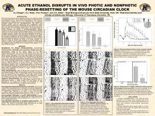
INTRODUCTION Alcoholism and alcohol abuse markedly disrupt brain systems closely and reciprocally tied to the circadian timing system located within the suprachiasmic nucleus (SCN) of the hypothalamus. The circadian timing system is regulated by photic and nonphotic input, mediated by glutamate release from the retinohypothalamic tract (RHT) and serotonin release from the raphe-SCN tract, respectively. Acute alcohol-induced perturbations of glutamatergic and serotonergic tonus within the SCN and subsequent impairments of photic and nonphotic phase-resetting of the circadian timing system are several folds: (1) alcohol increases the density of N-methyl-aspartate-D (NMDA) receptors through which glutamate binds, (2) alcohol inhibits glutamate release at the highly-ethanol sensitive NR2B subunit, and (3) alcohol increases serotonin release. Dose and time-dependent disruptions of circadian locomotor activity may secondarily contribute to an acute alcohol-induced impairment of phase-resetting. Given this information, the following experiments will be executed: 1) characterize ethanol pharmacokinetics (absorption, distribution, metabolism, and excretion) within the SCN, 2) observe the effects of an acute, systemic (i.p) injection of EtOH on photic phase-resetting, and 3) the effects of i.p. injections of EtOH and subsequent 5HT1A/7 agonist(+)-8-OH-DPAT on nonphotic phase-resetting of the circadian timing system in the male C57BL/6J mouse. ACUTE ETHANOL DISRUPTS IN VIVO PHOTIC AND NONPHOTIC PHASE-RESETTING OF THE MOUSE CIRCADIAN CLOCKA.J. Brager1, C.L. Ruby1, R.A. Prosser2, and J.D. Glass1. 1Dept Biological Sciences, Kent State University, Kent, OH, 2Dept Biochemistry and Cellular and Molecular Biology, University of Tennessee, Knoxville, TN. Figure 3: Pharmacokinetic profiles of acute, systemic ethanol injection. A response to 2.0, 1.0, or 0.5 g/kg is shown for mice outfitted with a microdialysis probe stereotaxically aimed at the lateral margin of the SCN. Independent of dose, ethanol concentrations peaked 40 min post-injection (a=2.0 vs. 1.0 and 0.5 g/kg; p<0.05; b=1.0 vs. 0.5 g/kg; p<0.05). METHODS Microdialysis For Ethanol Pharmacokinetics. Concentrically designed probes constructed from a 26-gauge stainless steel outer cannula and inner 12kDa MW cutoff hemicellulose dialysis tubing were stereotaxically aimed at the lateral margin of the SCN (anteroposterior=-0.046 mm from bregma; lateral=+0.02 mm from midline; horizontal=5.5 mm from dura), and secured to the skull with dental cement. Placement of the probe was verified from 60µm-thick frozen sections stained with cresyl violet. Probe efficiency is estimated to be ~10%. Two days post-surgery, the probes of freely-behaving mice were continuously perfused with filtered artificial cerebrospinal fluid (ACSF). Dialysate samples were collected every 20 minutes for 1 hour to measure baseline levels of ethanol. Following baseline sampling, mice received an i.p. injection of 2.0 g/kg, 1.0 g/kg, or 0.5 g/kg ethanol (diluted to 20% in physiological saline; n=3 per group), and dialysate samples were collected every 20 minutes for 5 hours after the injection. Samples were stored at -20°C and analyzed with an Analox AM1 (measures the amount of oxygen needed to convert ethanol to acetoldehyde). Photic Phase-Resetting. Male C57BL/6J mice entrained to a 12:12 L:D photocycle (LD) received an i.p. injection of 2.0 g/kg, 1.0 g/kg, 0.5 g/kg ethanol (diluted to 20% in physiological saline) or saline vehicle (n=7 per group) 30 min preceding a 30 min light pulse (25 lux) delivered from ZT16-16.5. Phase-shifting was measured by release into constant darkness (DD) using an Aschoff Type II procedure. Nonphotic Phase-Resetting. Mice entrained to LD received an i.p. injection of 2.0 g/kg, 1.0 g/kg, 0.5 g/kg ethanol (diluted to 20% in physiological saline) or saline vehicle (n=7 per group) 30 min preceding an i.p. injection of 10 mg/kg (+)-8-OH-DPAT at ZT6. Animals were released into DD to measure phase-resetting. * RESULTS Dose-dependent EtOH pharmacokinetics in the SCN. Time of peak concentration was independent of dose and occurred 40 minutes post-injection. The half life and peak concentration of absorbed EtOH were dose-dependent; a half life of 1.77 +/- 0.20 hr, 1.10 +/- 0.5 hr, and 0.58 +/- 0.08 hr were observed in 2.0 g/kg, 1.0 g/kg, and 0.5 g/kg, respectively with peak concentrations reaching 115.33 +/- 11.78 mM, 40.33 +/- 6.06 mM, and 23.00 +/- 5.77 mM, respectively (p<0.01). Dose-dependent attenuation of photic phase-resetting. Greatest attenuation was produced by the highest dose (2.0 g/kg) of EtOH with phase delays being about ~50% of vehicle (-0.7 +/- 0.2 h vs. -1.4 +/- 0.2 h, respectively; p<0.03). Dose-dependent attenuation of nonphotic phase-resetting. The highest dose depressed serotonergic phase advances (0.0 +/- 0.0 h; p<0.01). The extent of serotonergic phase advances did not differ between vehicle and thelower doses of EtOH (~0.5 h). Figure 4: SCN perfusion of ACSF or 500 mM of ethanol into the lateral margin of the mouse SCN. Perfusion commenced30 min preceding a 30 min (25 lux) light pulse or sham pulse (0 lux) at ZT14, using reverse microdialysis. Immediately following a light or sham pulse, mice were released into constant darkness to determine the magnitude of photic phase-resetting using the Aschoff Type II procedure. CONCLUSIONS 1. Ethanol pharmacokinetics in the SCN are dose-dependent. In all doses, Tmax coincides with the 30 min light pulse and (+)-OH-DPAT administrations in the photic and nonphotic phase-resetting experiments, respectively. 2. Ethanol attenuates photic phase delays in a dose-dependent manner with the highest dose having the most pronounced effect. 3. Ethanol attenuates serotonergic phase-advances in a dose-dependent manner with the highest dose having the most pronounced effect. Figure 2:Dose-dependent attenuation of nonphotic ((+)-OH-DPAT )phase-resetting. The protocol utilized to assess ethanol-induced impairments (above) and the dose-dependent attenuation of nonphotic phase-resetting (middle), and representative actograms of a mouse treated with 2.0 g/kg ethanol (top) or saline vehicle (bottom) and subsequent (+)-OH-DPAT are represented. Figure 1:Dose-dependent attenuation of photic phase-resetting. Schematics illustrate the protocol undertaken to assess ethanol-induced impairments (above) and dose-dependent attenuation of photic phase-resetting (middle), and representative actograms of a mouse treated with 2.0 g/kg ethanol (top) or saline vehicle (bottom). Acknowledgements: NIH AA015948 grant to R.A.P and J.D.G


