
Pediatric Case Study: Idiopathic Interstitial Lung Disease Presentation
A 13-year-old male presents with dyspnea on exercise, CT showing interstitial lung disease, and progressive pulmonary fibrosis. Treatment and follow-up are discussed.

Pediatric Case Study: Idiopathic Interstitial Lung Disease Presentation
E N D
Presentation Transcript
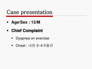
Case presentation • Age/Sex : 13/M • Chief Complaint • Dyspnea on exercise • Onset : 내원 3~4개월전
Present Illness • 내원 3~4개월 전부터 평소에는 괜찮으나 심한 운동시 숨이 차는 증상 있었으며, 내원 20일전 학교에서 실시한 흉부방사선촬영상 이상소견(결핵의심)보였으며, 내원 1주일 전 모대학병원에서 시행한 chest CT 상 interstitial lung disease 의심되어 조직검사 권유 받고 본원으로 전원 됨
Family History Tb contact (-) 집주변 공장 or 화학약품 노출 (-) Smoking exposure (-) Allergy F.Hx (-) 23 18
Past Medical History • G.A 40주, B.wt 3.2kg, FSVD • frequent URI Hx (-) • allergy Hx (-) • previous healthy child
Review of systems • Well-being appearance, weight loss(-) • Fever(-) • Cough/ Sputum/ Rhinorrhea(-/-/-) • tachypnea (-) • Dyspnea on exercise(+) : no interval change • Anorexia/ Nausea/ Vomiting(-/-/-) • Diarrhea/ Constipation(-/-) • Abdominal pain(-) • Dysuria/ Frequency/ Hematuria(-/-/-)
Physical examination I • Vital sign BP: 125/70 mmHg HR: 78 회/분 RR: 20 회/분 BT: 36.0℃(A) • Body Gauge Wt. : 33.2 kg (10-25percentile) Ht. : 146.7 cm (10-25 percentile) • General appearance not so ill looking appearance alert mental status • HEENT not anemic conjunctiva anicteric sclera PI (-)/ PTH (-) oral mucosa/ lip/ tongue : no focal lesion no palpable cervical LN
Physical examination II • Chest symmetric chestwall expansion CBS on BLF crackle (-), wheezing(-) chestwall retraction (-) RHB s murmur • Abdomen Soft and flat Normoactive B/S Liver/spleen : not palpable • Back and extremity Deformity (-) LOM (-) Swelling (-) Peripheral circulation : good • Skin rash (-)
Initial Lab Finding (06.07.03) • CBC 8970-14.2-310K ( 57.4 – 35.6 ) • CRP 0.08 • AST/ALT 33/26 • Ig G/A/M/E 1377/407/212/50.3 • AFB stain & culture (-), Tb-PCR (-),PPD (-) • ANA(-) • PFT : FVC 1.72L (61% pred), FEV1 1.47L (57% pred), FEV1/FVC 85% mild restrictive pattern
Imaging study Chest PA Ground-glass opacity with reticular opacity in the both lungs (reticulation is more prominent in the both basal lungs) Compatible with pulmonary fibrosis.
Chest CT: Lower lung zone 그리고 upper lung zone의 anterior lung으로 ground-glass opacities, reticulation의 pulmonary fibrosis .
Posterobasal segment of right lower lobe: End stage fibrosis (honeycombing) withlymphocytic infiltration
Aanterior segment of right upper lobe; Irregular parenchymal and subpleural fibrosis Interstitial diffuse infiltration of lymphocytes and patchy aggregation
Irregular subpleural and interlobular septal fibrosis with lymphocytic infiltration, and honeycombing (RLL)
Irregular subpleural and interlobular septal fibrosis with lymphocytic infiltration
Irregular subpleural and interlobular septal fibrosis with lymphocytic infiltration
Irregular subpleural and interlobular septal fibrosis with lymphocytic infiltration
Irregular subpleural and interlobular septal fibrosis with lymphocytic infiltration Patchy aggregation of lymphocytes
Irregular subpleural and interlobular septal fibrosis with lymphocytic infiltration:UIP pattern
Treatment • PD 2mg/kg (06.07.14 ~ 07.12.15) 1mg/kg (06.12.15 ~ 07.02.12) tapering .. stop Tx. (07.03.19) • Clarithromycin : anti-inflammatory dose (07.04.20 ~ 07.05.22) • PD 1.5 mg/kg (07.05.23~) Exercise intolerance (-), PFT : improving No Sx., Chest CT : aggr. Exercise intolerance (+), CXR : aggr.
F/U study • PFT • Chest CT (07.04.11) • 양측 upper lungs의 anterior subdural portion과 basal lung의 subpleural portion으로 honeycombing을 보이는 lesion의 내부 cyst의 size가 증가하였음. 이러한 lesion의 extent는 apex쪽으로 조금 증가한 양상임. • 비교적 preserve된 middle lung zone에는 새로 생긴 lesion 없음. Interval progression involving both upper and basal lungs.
Problem lists • Idiopathic Interstital lung disease • Progressive state • Cellular and fibrosing insterstitial penumonia (UIP pattern) • Subpleural and septal fibrosis and inflammatory cell infiltration, lesional variatiation, and pathchy lymphocytic infiltration, and ample of normal lung • End-stage fibrosis in RLL basal area (honeycombing)
ReviewInterstitial lung disease SMC PED Young-Hee Chung
Introduction • A group of rare, mostly chronic pulmonary disorders • High morbidity and mortality • More often in adults and is rare in children • Prevalence of PILD 0.36/100,000 • Pediatric interstitial lung disease (PILD) is difficult to define due to the diverse nature of ds. • No classification scheme for PILD is entirely satisfactory • Diffuse lung infiltrates and disordered gas exchange • Characterized by tachypnea, crackles, hypoxemia
Classification(1) • ILD can be of known or unknown etiology, or associated with other conditions, depending on whether the initiating inflammatory event has been identified • Approximately 50% of chronic ILD in children, no specific etiology is found • 1975 Liebow • the first to critically examine the pathological classification of ILD divided into 5 groups based on histological criteria • usual interstitial pneumonia (UIP), desquamative interstitial pneumonia (DIP), bronchiolitis obliterans with interstitial pneumonia (BIP), lymphoid interstitial pneumonia (LIP) and giant-cell interstitial pneumonia (GIP).
Classification(4) Pediatric pulmonology, 2004
Usual interstitial pneumonia (UIP)(1) • Pathologic pattern with clinical idiopathic pulmonary fibrosis or cryptogenic fibrosing alveolitis • Key histologic features : architectural destruction, fibrosis often with honeycombing, scattered fibroblastic foci, patchy distribution, involvement of the periphery of the acinus or lobule. • Fibroblastic foci : leading edge of the fibrotic process : essential to the diagnosis of UIP
Usual interstitial pneumonia (UIP)(2) • usually fails to respond to corticosteroid treatment • The prognosis of adultswith UIP is poor with the majority of patients succumbing within 5 years of diagnosis • In contrast, children given a diagnosis of idiopathic pulmonary fibrosis or cryptogenic fibrosing alveolitis often live much longer and have a nonprogressive course suggesting that they do not have UIP
Nonspecific interstitial pneumonitis (NSIP) • Histologically is a mixture of inflammation and fibrosis in the alveolar walls subdivided into a cellular and a fibrosing pattern • Differential diagnosis form UIP: Temporal uniformity of the alterations, the absence of honeycombing and the lack of active fibrosis • Cellular histologic pattern: have a better response to steroid therapy , may resolve completely • Fibrosing histologic pattern: likely to progress to end-stage lung disease. • better prognosis than UIP
Diagnosis • identifying the potential cause of the disease • establishing the histological picture for diagnostic, therapeutic, prognostic implication.
Chest X-ray • not prove sensitive • abnormal chest X-ray findings have been reported in almost all patients • 4 radiographic pattern : reticular, reticulonodular, reticulo-granular, ground-glass patterns
High-Resolution Computed Tomography • the extent and distribution of parenchymal disease and to select appropriate biopsy sites. • increase the level of diagnostic confidence for the diagnosis of pediatric ILD, improve diagnostic accuracy • Lynch et al. 1999, In a recent study of 20 children with biopsy-proven ILD, 56% of the confident first-choice diagnoses on HRCT were correct.
High-Resolution Computed Tomography • 5 distinct groups based on dominant HRCT features • (1)Airway disease (n = 5) (bronchiolitis obliterans or bronchocentric granulomatosis) showed geographic hyperlucency on CT. • (2)Septal disease (n = 4) (lymphangiomatosis, hemangiomatosis, or microlithiasis) showed septal thickening. • (3)Infiltrative lung disease (n = 7) (desquamative interstitial pneumonitis, hypersensitivity pneumonitis, or lymphoid interstitial pneumonitis) showed ground-glass opacity. • (4)Air-space disease (n = 3) (aspiration, vasculitis, or bronchiolitis obliterans organizing pneumonia) showed lung consolidation. • (5) Langerhans' histiocytosis (n = 1) showed cysts and nodules
Lung Biopsy • Gold standard for most types of PILD. • Open lung biopsy (OLB) or videoassisted thoracoscopic surgery (VATS) for obtaining tissue adequate for Dx • Fan et al, In a prospective study in a small group of immunocompetent children with ILD, • diagnostic yield was comparable for OLB and VATS • but the morbidity from VATS was clearly lower with respect to duration of surgery, chest tube, and hospitalization.
Treatment (1) Elimination of the etiological agent : infectious, environmental, drugs, irradiation, etc (2) Therapy of associated disease (3) Corticosteroid therapy (oral or i.v. [boluses of methyl prednisolone]) + hydroxychlroquine in idiopathic ILD (4) Lung transplantation in severe cases
Corticosteroid • Prednisolone • used in initial once daily doses of 1-2 mg/kg/day, for 6 to 8 weeks gradually tapered off • depending on the clinical response and the improvement in respiratory function • Treatment with oral steroids can be continued for years. • Methylprednisolone IV pulse • currently prefer this method to oral daily or every-other-day therapy because of fewer side effects. • 10-30 mg/kg/day boluses, with a maximum of 1g i.v over 1hr, for 3 consecutive days at intervals of one month, for a total of 6 months or more.
Chloroquine, its derivative, Hydroxychloroquine • They are primarily anti-fibrotic but can be effective in patients who have failed to respond to oral or i.v corticosteroids. • Previously, chloroquine was widely used for this condition but an important side-effect was retinopathy. Hydroxychloroquine is now used routinely, as the incidence of ophthalmic problems with this agent is much less. • Current practice is to carry out a baseline ophthalmology review before starting treatment and then review at 2-yearly intervals if treatment is continued throughout this time.
Other therapy • Colchicine • powerful inhibitor of fibroblast protein Synthesis • used to suppress the release of growth factors • Cyclophosphamide , other immunosuppressants (azathioprine, methotrexate) • depress lymphocytic function and reduce the number of neutrophils. • significant improvements in the short-term, their long-term efficiency is not yet known. • Newer therapies • directed against certain cytokines, oxidants, and growth factors that may be involved in the fibrotic process hold promise for the future. • Lung transplantation
Prognosis • These treatments result in a successful outcome in up to 65% of cases • The overall mortality is 15%. • Useful measure of outcome : Of the clinical features present at time of initial evaluation, crackles, clubbing, family history of ILD, and symptom duration were not associated with decreased survival, but the severity of illness classification
Radiological versus histological diagnosis in UIP and NSIP: survival implications(1)Thorax 2003;58:143-8
Radiological versus histological diagnosis in UIP and NSIP: survival implications(2)Thorax 2003;58:143-8
Radiological versus histological diagnosis in UIP and NSIP: survival implications(3)Thorax 2003;58:143-8 NSIP (HRCT+Bx.) NSIP (HRCT)+UIP (Bx.) UIP (HRCT+Bx.)
Radiological versus histological diagnosis in UIP and NSIP: survival implications(4)Thorax 2003;58:143-8 • (1) patients with an HRCT pattern of UIP are likely to have a histopathological pattern of UIP, but patients with an HRCT pattern other than UIP may have either a histological pattern of UIP or NSIP on the surgical lung biopsy specimen • (2) HRCT features add prognostic information to the histological diagnosis of UIP: survival was worse if patients with histological UIP had an HRCT picture felt by expert radiologists to be definite or probable UIP compared with patients with histological UIP but an atypical HRCT picture for UIP • (3) HRCT has limited specificity in identifying histological NSIP.
Usual interstitial pneumonia and non-specific interstitial pneumonia :serial thin-section CT findings correlated with pulmonary function(1)Korean Journal of Radiology; 2005 September; 6(3):143-152 • To demonstrate and compare the serial high-resolution CTs (HRCT) and the pulmonary function test (PFT) findings of the usual interstitial pneumonia (UIP) and the non-specific interstitial pneumonia (NSIP). • 35 patients having UIP without significant honeycombing (UIP-w/o hc, < 5% of honeycombing at CT), 35 patients having UIP with honeycombing (UIP-w/i hc, ≥ 5% of honeycombing), and 25 patients with NSIP
Usual interstitial pneumonia and non-specific interstitial pneumonia :serial thin-section CT findings correlated with pulmonary function(2)Korean Journal of Radiology; 2005 September; 6(3):143-152 • On the initial CT, significant differences were present between the UIP-w/i hc patients and both the UIP-w/o hc patients and the NSIP patients in the overall extent, ground-glass opacity (GGO) away from the reticulation, reticulation and honeycombing (all p < 0.05) • Five (14%) of the 35 patients with UIP-w/o hc, 16 (46%) of the 35 patients with UIP-w/i hc and three (12%) of the 25 patients with NSIP died (p = 0.002, comparison for the three groups). • On CT, NSIP and UIP-w/o hc patients have similar patterns of parenchymal abnormalities and a similar likelihood of change in the extent of disease on follow-up. Patients with UIP-w/i hc have distinctive features and a worst prognosis
소아의 간질성 폐질환에서 폐 조직생검 소견과 임상 양상(1)소아과 2002년 45권 1호 79-87 • 1990년 1월부터 1999년 8월까지 서울중앙병원 소아과에서 개흉폐생검으로 간질성 폐질환으로 진단된 15례와 호흡곤란이나 반복적인 천명 등 호흡기 증상과 HRCT상에서 간질성 폐질환의 특징적 소견이 관찰되어 폐색성 세기관지염으로 진단된 6례 등 총 21명의 환아들을 대상으로 후향적으로 추적 관찰하여 분석하였다 • 1년 5개월(범위, 1개 월-7년 5개월) • 고해상도 흉부단층촬영 상 mosaic perfusion(12례)이 가장 많은 빈도로 관찰되었고, bronchial wall thickening(9례), bronchiectasis(8례), multiple segmental atelectasis (5례), ground glass opacity(5례)
소아의 간질성 폐질환에서 폐 조직생검 소견과 임상 양상(2)소아과 2002년 45권 1호 79-87 • 개흉폐생검술을 시행한 15례 중에서 14례(93.3%)에서 조직학적으로 확진이 가능하였고 간질성 폐장염이 6례(NSIP 3례, DIP 2례, UIP 1례)로 가장 많았으며 폐섬유증 2례, CMV폐장염 2례, 미만성 흡인성 세기관지염 1례, 폐림프관종증 1례, BOOP 1례, 폐 조직구증식증 1례로 진단 • 총 21례 중 15례에서 methylprednisolone pulse 요법을 행하였고, 그 방법은 methylprednisolone 30 mg/kg/일을 2일 간격으로 6회 정주하고 그 후로 1개월 간격으로 12회 주사 • Methylprednisolone pulse치료를 시행한 15례 중 1례는 사망하였고 1례에서는 점수(Denver protocol, 1997) 변화가 없었으나 13례(86.6%)에서는 임상 점수가 호전되었다.
Idiopathic interstitial pneumonitis in children :A national survey was carried out in the United Kingdom and IrelandPediatr Pulmonol. 2002;34:23–29. • over a 3-year period • 46 cases were identified, 29 males and 17 females. • Conventional treatment with pulsed methylprednisolone, prednisolone, or hydroxychloroquine, singly or in combination, resulted in an excellent response in 65% of cases. Seven children died (15%).


