
Preface
Preface. All cytoskeletal elements known from eukaryotic cells are also present in bacteria, where they perform vital tasks in many aspects of the physiology of the cell.

Preface
E N D
Presentation Transcript
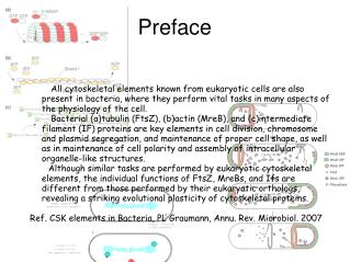
Preface • All cytoskeletal elements known from eukaryotic cells are also present in bacteria, where they perform vital tasks in many aspects of the physiology of the cell. • Bacterial (a)tubulin (FtsZ), (b)actin (MreB), and (c)intermediate filament (IF) proteins are key elements in cell division, chromosome and plasmid segregation, and maintenance of proper cell shape, as well as in maintenance of cell polarity and assembly of intracellular organelle-like structures. • Although similar tasks are performed by eukaryotic cytoskeletal elements, the individual functions of FtsZ, MreBs, and Ifs are different from those performed by their eukaryotic orthologs, revealing a striking evolutional plasticity of cytoskeletal proteins. • Ref. CSK elements in Bacteria, PL Graumann, Annu. Rev. Microbiol. 2007
Observe how cell morphology affects bacterial cell division, • and the effects of specific bacterial CSK protein deficiencies. • • Explain division patterns via : • +observing the perfect globular YLS2 strain (∆mreB) • +how it contrasts with MC1000(wild type) • +using YLS1(∆minCDE) for more explicit observation of • the Min system Aim
YLS1 YLS2 MC1000 YLS1 YLS2 Growth Chart MreB deficient Wildtype MinCDE deficient Property 1 esp. mini cells: highly motile 3 Only during some division periods 2 Motility Preparation: Temperature (Celsius) 30 30 35 Incubation hours 3 3~3.5 3.5~5 before microscopic observations 1
Summary (a) Exterior forces, altering cell morphology, may effect and even determine division sites. it may possibly be attempted by using something in nano scale to make more desirable and controllable way of interference (b) Via the YLS2 DIC images, it clearly suggests that the min oscillation diverts 90o during sometime within the last stages of cell division. should be observed and assisted in the future with fluorescent methods • To be improved: (a) staining the membrane with clear visual outcomes (b) all-time steady focus when recording • Future work (a) More explicit observations on DIC images (b) Approach on Fluorescent Microscopy analysis in 2D and 3D
PLUS! Loads and Loads of Thanks to Everyone~