Advanced Multimodal Medical Visualization Techniques
Explore cutting-edge visualization methods such as RT3D, virtual endoscopy, image fusion, and more for precise medical imaging applications. Learn about quantification of tumor volumes, aneurysms, calcium scoring, and cardiac dynamics with virtual dissection capabilities. Dive into the assessment of aortic aneurysms and Larnygo-Tracheal stenosis with advanced segmentation and skeletonization techniques. Develop expertise in using Active Contour Models (Snakes) for vessel analysis and Fuzzy Connectedness for precise airways imaging. Take your medical imaging skills to the next level with this comprehensive guide.

Advanced Multimodal Medical Visualization Techniques
E N D
Presentation Transcript
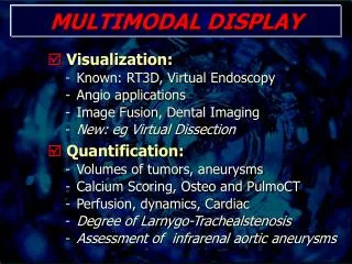
MULTIMODAL DISPLAY • Visualization: • Known: RT3D, Virtual Endoscopy • Angio applications • Image Fusion, Dental Imaging • New: eg Virtual Dissection • Quantification: • Volumes of tumors, aneurysms • Calcium Scoring, Osteo and PulmoCT • Perfusion, dynamics, Cardiac • Degree of Larnygo-Trachealstenosis • Assessment of infrarenal aortic aneurysms
Prerequisites • Segmentation • (Region Growing) • Fuzzy Connectedness (air filled organs) • Active Contour Models (vessels) • snakes, ballons • Centerline of tubular objects • Skeletonisation
Seg – Fuzzy Connetedness Udupa et al., Graphical Models and Image Processing,l 1996
Fuzzy Connectedness - Performance • Airways (200-300 slices) • Incorrect slices – manuell orrection: • Normals: n=2.1 • Pathologic: n=3.9 • Time: 15 – 20s p=0.06 n.s.
Seg – ACM - Snakes • Assumptions: • Vessels round -> elliptic • Only minimal changes from slice to slice • 2.5D approach: • Set of slices initialized • For every slice: • Contour of prev.slice used for starting • Snakes adopt contour according forces in x and y-direction
Skeletonisation The skeleton of an object is the locus of the centers of all the maximal inscribed hyper-- spheres