Download
1 / 45
490 likes | 1.53k Vues
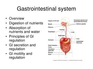
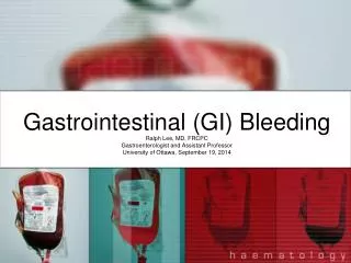
Gastrointestinal (GI) Bleeding. Ralph Lee, MD, FRCPC Gastroenterologist and Assistant Professor University of Ottawa, September 19, 2014. Objectives. Define upper and lower gastrointestinal (GI) bleeding. Outline the etiologies and clinical features of upper and lower GI bleeding

E N D
Gastrointestinal (GI) Bleeding Ralph Lee, MD, FRCPC Gastroenterologist and Assistant Professor University of Ottawa, September 19, 2014
Objectives • Define upper and lower gastrointestinal (GI) bleeding. • Outline the etiologies and clinical features of upper and lower GI bleeding • Apply a systematic clinical approach to GI bleeding. • Outline the investigation and management of GI bleeding • Recognize clinical indicators suggesting urgent versus non‐urgent assessment.
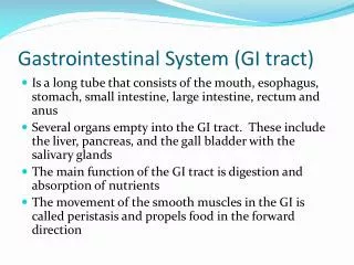
What is Gastrointestinal Bleeding? (1) • Bleeding from anywhere in the GI tract • Esophagus to rectum • Traditionally, classified into two groups, based on presumed location of bleeding: • Upper • Lower • 3rd category, ‘Mid- GI’ bleeding, is present, but infrequently used
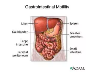
What is Gastrointestinal Bleeding? (2) • Upper GI (UGI) Bleeding • Traditional: Proximal to the Ligament of Treitz • New: Proximal to the Ampulla of Vater • Lower GI (LGI) Bleeding • Traditional: Distal to Ligament of Treitz • New: Distal to terminal ileum • Mid GI bleeding • New: Ampulla of Vater to terminal ileum
UGI Bleeding • 75 – 80% of GI bleeding • annual hospitalization rate = 160/100,000 (US) • Mortality rate = 3.5 – 10% • Sex – M:F – 2:1 • incidence with age • More likely to present with hemodynamic instability due to rich blood supply of UGI tract
UGI BleedingCauses (Common) • Peptic Ulcer disease (20 - 50%) • Varices (5 - 20%) • AV Malformations (5%) • Mallory-Weiss tears (8 - 15%) • Tumours (5%) • Erosions (8 - 15%) • Dieulafoy’s lesions (1%)
UGI BleedingCauses (Other) • Other (11%) • Gastric Antral Vascular Ectasia (GAVE) • AKA ‘Watermelon’ stomach • Portal hypertensive gastropathy • Hemobilia • Hemosuccus pancreatitis • Aortoenteric fistulas • Cameron’s lesions/ulcers
UGI BleedingHow Do They Present? (1) • Hematemesis • Vomiting blood • Bright red blood, clots, ‘coffee ground’ emesis • Melena • Passage of black, tarry, foul-smelling stools • Digested blood • Appears with ≥ 50cc of bleeding from UGI tract • Things that mimick melena: • Iron pills - greenish • Bismuth subsalicylate (PeptolBismol™) - non-foul smelling
UGI BleedingHow Do They Present? (2) • Bowel movement frequency • Blood is cathartic and a great laxative • Rough indicator of rapidity of bleeding • Hemodynamic symptoms • Pre/Syncope • Orthostatic dizziness/lightheadedness • Chest pain, dyspnea
UGI BleedingHow Do They Present? (3) • (Hematochezia - red blood per rectum) • Usually sign of LGI Bleeding, but can occur with rapid UGI bleeding (i.e. ≥ 1000cc) • patient usually hemodynamically unstable • Other symptoms • Dependent on cause • Abdominal pain, heartburn, dysphagia, nausea, vomiting
LGI Bleeding • 20 – 30% of gastrointestinal bleeding • Usually less hemodynamically significant, higher Hb level, less blood transfusion requirements than UGI bleeds • Increased incidence with age • Mean age at presentation: 63 – 77 • Mortality rate among hospitalized acute lower GI bleeds – 2 - 4%
LGI BleedingCauses • Diverticulosis (25 - 65%) • Cancers/polyps (17%) • Colitis/ulcers (18%) • IBD, ischemic, vasculitis, infectious, radiation-induced, NSAID-induced • Unknown (16%) • Angiodysplasia(3 - 15%) • Other (8%) • Post-polypectomy, stercoral ulcers, aorto-colonic fistulas • Anorectal(24 - 64%) • Fissures, hemorrhoids
LGI BleedingHow Do They Present? (1) • Red Blood per Rectum • Bright Red Blood Per Rectum (BRBPR) – left colonic? • Dark/maroon – right colonic/lower small bowel? • Stool • frequency – blood is cathartic • form – diarrhea • Location of blood • Surface/side of stool – perianal source? • Mixed in stool – R colonic? • (Melena)* • Usually sign of UGI bleeding, but can occur with distal small bowel, cecum or R-sided colonic bleeding source • Typically, hemodynamically stable with less rapid bleeding
LGI BleedingHow Do They Present? (2) • Other Symptoms • Dependent on cause • Fecal urgency, tenesmus, incontinence • Abdominal pain/cramps • Fevers/chills • Weight loss
GI BleedingOther important historical items • Past medical history: • GI • Previous PUD/H. Pylori infection? • GI Malignancy? • Previous endoscopies • Previous polypectomy • Diverticulosis • IBD • Cirrhosis? • Varices? • Cardiac • CAD, angina, MI, CHF • Previous aortic aneurysms/grafts/vascular surgery? • Previous radiation therapy? • Bleeding episodes • Medications • Anti-platelet agents (i.e. ASA, Plavix), anticoagulants (i.e. warfarin, pradaxa) • NSAIDs • Habits – EtOH • Family history: • PUD, gastric cancer • Colonic polyps, cancer • IBD
GI BleedingPhysical Exam (1) • Look for: • Hemodynamic instability • Intravascular depletion • Potential etiologies of bleeding • General: • Altered mentation, jaundice • Vitals • Hypotension • Tachycardia • Orthostatic changes • in BP and/or HR with position change from supine to standing • Suggests intravascular volume depletion of ≥ 2L • Head and Neck: • Conjunctival pallor • Scleral icterus • Dry mucous membranes, furrowed tongue, JVP • Chest: • skin turgor at sternal angle • Axilla: dry or moist • Abdomen: • Tenderness, masses, hepatosplenomegaly, stigmata of chronic liver disease • Digital rectal exam: • Red blood? Melena? • Hemorrhoids, fissures • Masses
GI BleedingManagement (Overview) • ABC’s • Resuscitation • Focused History and Physical Exam • Upper vs. lower GI bleed • Investigations • Treatment
GI BleedingInitial Management • ABC’s • Resuscitation • Intravenous (IV) access • Monitoring • Need for monitored area ? (i.e. ICU) • Cardiac, respiratory (i.e pulse oximetry) • Volume re-expansion • IV fluids • Keep patient NPO (nil per os) • Focused history and physical exam • Upper vs. lower GI Bleed
GI BleedingInvestigations (1) • Bloodwork • Type and crossmatch - PRBC’s, blood products • Complete Blood Cell Count • Hemoglobin • MCV – mean corpuscular volume • MCV Fe deficiency chronic blood loss? • Platelets • Clotting ability • Electrolytes
GI BleedingInvestigations (2) • Blood Urea Nitrogen (BUN), Creatinine • Intravascular dehydration renal perfusion BUN, Cr • UGI bleed blood digestion in stomach/duodenum protein absorption urea nitrogen • [BUN x 10]: Cr > 1.5:1 May suggest UGI bleed • Liver enzymes and liver function tests • INR - Coagulation status (i.e. INR) • Liver disease (i.e. cirrhosis) • Albumin • INR • AST > ALT • Platelets, MCV • (Nasogastric aspirate)
GI BleedingInitial Treatment (1) • Fluid Management • IV crystalloids (i.e. normal saline); colloids (i.e. volume expanders) • Transfusion - Packed red blood cells • Reverse anti-coagulation • INR - Vitamin K, fresh frozen plasma, prothrombin complex concentrate (i.e. Octaplex) • Platelets – Platelets transfusion
GI BleedingInitial Treatment (2) • Pharmacotherapy • UGI Bleeds: • IV Proton Pump Inhibitor • Mechanism: pH > 6 Promotes clot stability • acid/pepsin on lesion • platelet aggregation + fibrin formation • Empirically started to treat possible PUD until EGD performed • IV PPI before endoscopy: • Lau et al. (2007): need for endoscopic therapy + accelerated healing of ulcers • Cochrane Meta-analysis (Sreedharan et al., 2010): • DOESN’T: mortality, rebleeding or progression to surgery • DOES: high risk stigmata, need for endoscopic therapy • IV PPI after endoscopic therapy: rebleeding, mortality • Dose: Pantoprazole 80mg IV bolus, then 8mg/h x 72 hours
GI BleedingInitial Treatment (3) • IV Somatostatin (i.e. octreotide) • For possible varicealbleeds (i.e. patients with cirrhosis) – started empirically • Mechanism: splanchnic vasoconstriction portal hypertension bleeding • Prokinetics (20 – 120 minutes before) • i.e. metoclopramide (Maxeran) or IV erythromycin • To clear UGI tract of blood for better visualization • Meta-analysis (Barkun et al., 2010): need for repeat endoscopy • LGI Bleeds: No specific medications
Urgent vs. Non-urgent Management Urgent Non-urgent Young, healthy, minimal bleeding • Hematemesis • Serious co-morbid illness • Malignancy, cirrhosis • Hemodynamic instability • Shock • SBP < 100mmHg • HR > 100bpm • (Orthostatic hypotension) • Hb < 80 • Transfusion requirement > 2u PRBC’s • Severe, ongoing bleeding
GI BleedingEndoscopy • UGI Bleed – EGD • Timing: Within 24 hours of presentation • To: • Diagnose cause of bleeding (high sensitivity/specificity) • Stratify risk of rebleeding/adverse events • Potentially treat underlying pathology • LGI Bleed – Colonoscopy • Timing: Controversial • Severe bleeds: Within 8 – 24 hours • Generally, LGI bleeds less severe than UGI bleeds • Often, more difficult to identify source; therefore, mostly diagnostic rather than therapeutic • Unclear: EGD Colonoscopy
Upper vs. Lower EndoscopyPresumed Upper GI Bleed Hematemesis EGD
Upper vs. Lower EndoscopyOccult Bleeding • Occult bleeding • Stool testing positive for occult blood • Unexplained Fe deficiency anemia
GI BleedingEndoscopic Hemostasis (1) • Injection • i.e. Vasoconstrictors (i.e. epinephrine), saline, sclerosants, tissue adhesives • Creates submucosal cushion of fluid which tamponades site +/- vasoconstriction • Thermal therapy • i.e. Mono/bipolar electrocoagulation, Argon Plasma Coagulation (APC), Laser Photocoagulation • Cauterizes vessel closed
GI BleedingEndoscopic Hemostasis (2) • Mechanical therapy • i.e. Hemoclips, rubber bands • Closes and tamponadesvessel • Animation • http://www.youtube.com/watch?v=59uO-8UVC2A • Hemospray • New therapy • Nano particle spray
UGI BleedingPeptic Ulcer Disease (1) • Most commonly due to NSAIDS, H. Pylori • 75 – 80% stop spontaneously • Ulcer appearance indicates risk of rebleedingand determines whether therapy required • Forrest Classification
UGI BleedingPeptic Ulcer Disease (2) • Endoscopic intervention: • Risk of rebleeding • Need for surgery • Mortality
UGI BleedingVarices • Often unstable UGI bleeds • Esophageal – Options: • Ligated with rubber bands (band ligation) • Injection with sclerosants • Gastric – Options: • Injected with ‘glue’ (cyanoacrylate) • Band Ligation • If endoscopy unsuccessful: • Transjugular intrahepatic portosystemic shunt (TIPS) • Liver transplant
LGI BleedingDiverticular Bleeding • Complicates 3 – 15% with colonic diverticulae • Pathophysiology: • Trauma of vasa recta at neck or dome of diverticulum • Presentation: • Painless hematochezia • Treatment: • 75 - 80% resolve spontaneously • Recurrence within 4 years • 25 – 40% • Endoscopic hemostasis • If site can be identified • < 30d rebleeding: Uncommon • Angiography (85% effective) • < 30d rebleeding: 22% • Surgical resection
LGI BleedingAngiodysplasia • Increases with age • Pathophysiology • Degenerative changes • Chronic, intermittent obstruction of submucosal vessels • Presentation • Usually asymptomatic • Overt or occult bleeding • Usually in right colon • Treatment • Iron replacement • Cauterization if bleeding or Fe deficiency anemia • (Estrogen/progesterone)
GI BleedingOther Options (1) • If endoscopy fails to identify source or fails to stop the bleeding • Mesenteric angiography • Performed by interventional radiology • Catheter introduced through femoral artery, passed to celiac trunk, SMA, IMA + branches to: • Diagnose bleeding site • Requires blood loss of ≥ 0.5-1mL/min • Low sensitivity (47%) but high specificity (100%) • Perform therapy • Feeding arteries can be embolized with microscopic gel foam, microcoilsor EtOH particles • Success rate: 52 – 94% • http://www.youtube.com/watch?v=jvi2WwvXIew • Complications (17%): Nephrotoxicity, bowel infarction, hematomas
GI BleedingOther Options (2) • RBC scan • Patient infused with technetium tagged RBC’s to locate site of bleeding • Disadvantages: • Not therapeutic • Only localizes bleeding to generalized area of abdomen • Requires blood loss ≥ 0.1mL/min • Higher sensitivity; lower specificity • Surgery • Intraoperative enteroscopy • Oversew, resection
Summary (1) Upper GI Bleed Lower GI Bleed Dark/bright red blood per rectum (hematochezia) Longer course Tend to be more hemodynamically stable • Hematemesis • Melena • Hemodynamic instability • Elevated BUN x 10: Cr
Summary (2) • PUD is most common cause of UGIB bleeding • Diverticular disease is the most common cause of significant LGIB • Approach includes • ABC’s • Resuscitation • History and Physical Exam • Initial Investigations • Initial Treatment • Endoscopy • Radiology • Surgery • IV PPI’s are started empirically if UGIB is suspected • Endoscopy is the primary diagnostic and therapeutic tool for GI bleeds