Detection of HCV Protein Expression and T Cell Responses in BALB/c Mice Using VEE/SIN Vector
This study investigates the expression of Hepatitis C Virus (HCV) proteins (E1, E2, NS3, NS4, NS5) using VEE/SIN replicon particles in BALB/c mice. Through Western blotting, we assess the expression profiles of these proteins, comparing results from cell lysates of mock-infected mice to those infected with VEE/SIN-NS345 control and recombinant protein controls. Additionally, we analyze cellular immune responses via intracellular cytokine staining (ICS) and FACS to evaluate CD4+ and CD8+ T cell activation following E1E2/MF59 immunization.

Detection of HCV Protein Expression and T Cell Responses in BALB/c Mice Using VEE/SIN Vector
E N D
Presentation Transcript
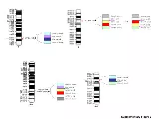
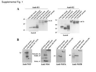
Supplemental Fig. 1 Anti-E1 Anti-E2 A VEE/SIN-E1E2P7 VEE/SIN-E1E2P7 r E1E2p7 protein) rE1E2P7 protein r E1E2P7 protein VEE/SIN-E1E2P7 VEE/SIN-E1E2P7 VEE/SIN-NS345 VEE/SIN-E1E2 VEE/SIN-E1E2 rE1E2p7 protein VEE/SIN-NS345 VEE/SIN-E1E2 VEE/SIN-E1E2 35 75 * 33-35 * * 68-74 25 50 35 15 25 EndoH EndoH B rSOD-NS5 (NS5A/B) rSOD-NS5 (NS5A/B) rSOD-C33C (NS3) rSOD-C100 (NS4) VEE/SIN-NS345 VEE/SIN-NS345 VEE/SIN-NS345 VEE/SIN-NS345 rE1E2p7 protein rE1E2p7 protein rE1E2p7 protein rE1E2p7 protein * * * * 70 NS4A/B: 35 * 69 * * NS4B: 27 * 57 * * NS4A: 8 Anti-NS3 Anti-NS4 Anti-NS5A Anti-NS5B
Legend of supplemental Fig. 1 Detection of HCV protein expression from VEE/SIN replicon particles by Western blotting. A. Detection of E1 and E2 expression from the BHK cell lysates with VEE/SIN-E1E2 and VEE/SIN-E1E2p7 infection. VEE/SIN-NS345 is a negative control. rE1E2p7 protein is a positive control. The size was indicated corresponding to E1 (33-35kDa) and E2 (68-74kDa). EndoH: protein was treated with EndoH (Biolab) at 37ºC for 1 hour. B. Detection of NS3, NS4, NS5A and NS5B expression from the cell lysates with VEE/SIN-NS345 infection. rE1E2p7 is a negative control. SOD-C33C (a.a. 1192-1457), SOD-C100 (a.a. 1569-1931), and SOD-NS5 (a.a. 2054-2995) are recombinant proteins used as positive controls for the detection of NS3, NS4, NS5A and NS5B respectively. The size was indicated corresponding to NS3, NS4A, NS4B, NS4A/B, NS5A and NS5B. * Indicated the specific detection of the proteins.
Supplemental Fig. 2 A B PBS (1,2,3) E1E2/MF59 (1,2,3) PBS (1,2,3) E1E2/MF59 (1,2,3) VEE/SIN-E1E2 (1,2,3) E1E2/MF59/CpG (1,2,3) VEE/SIN-E1E2 (1,2,3) E1E2/MF59/CpG (1,2,3) E1E2/MF59 (1,2) VEE/SIN-E1E2 (3) E1E2/MF59/CpG (1,2) VEE/SIN-E1E2 (3) E1E2/MF59 (1,2) VEE/SIN-E1E2 (3) E1E2/MF59/CpG (1,2) VEE/SIN-E1E2 (3) IFN-g IFN-g CD8 CD4
Legend of supplemental Fig. 2 Detection of CD4+ and CD8+ T cells responsesby ICS/FACS. BALB/c mice (n = 10) received three injections i.m. at 3-week intervals by the immunogens as indicated. The spleen cells were harvested 2 weeks after the last immunization and stimulated with 10 mg/ml of HCV specific peptides for ICS and FACS analysis. The data are presented as dot plots of ICS for CD4+ and IFN-g after E1 peptide pool stimulation (A) and for CD8+ and IFN-g after CD8 E2 peptide stimulation (B). Significant responses are indicated by circles in the upper-right quadrant.
Supplemental Fig. 3 * PBS (1,2,3) E1E2/MF59 (1,2); VEE/SIN-E1E2 (3) E1E2/MF59 (1,2,3) VEE/SIN-E1E2 (1,2); E1E2/MF59 (3) VEE/SIN-E1E2 (1,2); E1E2/MF59/CpG (3) E1E2/MF59/CpG (1,2); VEE/SIN-E1E2 (3) VEE/SIN-E1E2 (1,2,3) E1E2/MF59/CpG (1,2,3)
Legend of supplemental Fig. 3 VEE/SIN-E1E2 immunization and Prime/boost regimen with E1E2/MF59 and VEE/SIN-E1E2could stimulate good CD8+ T cells responses. BALB/c mice (n = 10) were received three injections i.m. at 3-week intervals by the immunogens as indicated. The spleen cells were harvested at 2 weeks after the last immunization and stimulated with 10 mg/ml of HCV specific peptides for ICS and FACS analysis. 20-mer over-lapping peptide pools for HCV-1a E1 region (E1 pool) and E2 region (E2 pool), and single peptide to E2 for CD4+ T cells (CD4 E2 pep) and CD8+ T cells (CD8 E2 pep) were used for stimulation. The data are presented as mean total percentages of CD8+IFN-g+ cells from two pools (five mice per pool). * P<0.05 as compared with PBS control group.