Download
1 / 62
640 likes | 1.11k Vues
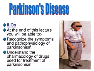
At the end of this participant you will be able to: Know the differences between ischemic and hemorrhagic stroke Recognize signs and symptoms of stroke Be able to use the Cincinnati Prehospital Stroke Scale Discuss major principles of prehospital assessment and treatment for acute stroke

E N D
At the end of this participant you will be able to: • Know the differences between ischemic and hemorrhagic stroke • Recognize signs and symptoms of stroke • Be able to use the • Cincinnati Prehospital Stroke Scale • Discuss major principles of prehospital assessment and treatment for acute stroke • Appreciate importance of rapid transport to Accredited Stroke Center 2
Appreciate importance of notifying ED before arrival (Calling Stroke Alert) • Discuss major principles of ED stroke care • Importance of rapid triage and early CT for stroke victims • Understand the potential use of thrombolytics (IV-tPA) for selected patients with acute ischemic stroke • Appreciate importance of rapid transport from an ED to an Accredited Stroke Center 3
Use of National Institutes of Health Stroke Scale (NIHSS) • Guidelines for managing hypertension in stroke patients • Clinical differences between ischemic and hemorrhagic stroke • Treatment differences between ischemic and hemorrhagic stroke • Appreciate importance of rapid transport from an ED to an Accredited Stroke Center 4
According to the American Heart Association stroke is the third leading cause of death in U.S. and leading cause of disability • Approximately 700,000 people each year will suffer from a stroke, either for the first time or with a history of stroke; Of those patients, approximately 158,000 will die as a consequence of the stroke. • One-third of strokes occur in patients younger than 65 years. • Men are at higher risk than women. • About 85% of strokes are ischemic in nature • About 15% of strokes are hemorrhagic in nature • EMS plays a large role as early recognition and treatment. This is key in reducing the mortality rates from strokes.
A stroke or Cerebral Vascular Accident (CVA) or “Brain Attack” is a neurologic deficit that causes a change in the patient’s ability to speak, feel, or move. • When these changes are noted, the EMT should recognize that something has affected the patient’s central nervous system. • This could be a medical or traumatic cause This power point will be limited to the presentation of a nontraumatic brain injury, or stroke.
Stroke • The symptoms that the patient presents with is a reflection of the area of the brain that has had a disruption of blood flow. • Most commonly, strokes affect the regions of the brain that control speech, sensation, and muscle function. • Paralysis, facial droop, monoplegia, hemiplegia, and speech disturbances are common findings.
Stroke is classified as hemorrhagic or ischemic and further subdivided by etiology • Ischemic stroke • Embolic • Thrombotic • Hypoperfusion • Hemorrhagic stroke • Intracerebral hemorrhage • Nontraumatic subarachnoid hemorrhage
Ischemic Stroke – This type of stroke is caused by a sudden occlusion of a blood vessel in the brain, a similar mechanism that is seen with a heart attack.
Hemorrhagic Stroke – This type of stroke occurs when a blood vessel in the brain bursts and allows blood to collect in or around the brain tissue.
In either instance, it is the lack of blood flow and oxygen that causes the dysfunction in the brain, and the accompanying signs and symptoms.
Nausea/vomiting • Dizziness, weakness • Headache • Impaired vision • Vertigo, tinnitus • Difficulty speaking, swallowing • Abnormal gait, weak extremities • Hemiparesis, quadriparesis • Sensory loss, seizures • Pupil abnormalities • Dilated • Constricted • Unreactive • Sluggish
“Hey you ….. Don’t spend a lot of time to determine the specific cause! Do Prehospital Clinical Assessment
Cincinnati Prehospital Stroke Scale (CPSS) • Assess for • Facial droop • Arm drift • Abnormal speech
Assess • Facial droop (have patient smile) • Normal: Both sides of the face move equally • Abnormal: One side of face does not move as well
Assess • Arm drift (have patient hold arms out for 10 seconds) • Normal: Both arms move equally or not at all • Abnormal: One arm drifts compared to the other, or does not move at all
Assess • Abnormal speech (Have the Pt. say) “you can’t teach an old dog new tricks” • Normal: Patient uses correct words with no slurring • Abnormal: Slurred or inappropriate words, or mute
If positive on one or all of the three tests Transport to the closest Accredited Stroke Center and call a STROKE ALERT
CHRISTUS Spohn Shoreline1-361-881-3811 600 Elizabeth St. Corpus Christi, Tx • CCMC Doctors Regional 1-361-761-1467 3315 S. Alameda Corpus Christi, TX • CCMC Bay Area 1-361- 761-3637 7101 S. Padre Island Dr. Corpus Christi, Tx
Time = Brain! Assessment and Treatment of a stroke patient by EMS can make a difference!
Scene evaluation • Initial assessment • Focused History “SAMPLE history/Vital signs/Check the blood glucose level” • Detailed Physical examination (as needed) • Ongoing Assessment • Treatment
Assessment: Scene Size-Up • Dispatch information may alert you to this emergency if there is knowledge of neurological deficits or altered mental status. • Look for evidence of trauma, drug use, or alcohol. • The patient’s clothing may indicate approximately when the symptoms started. • Call for backup if extrication from the residence will be difficult. • Remember to take BSI precautions.
Assessment: Initial Assessment • Establish mental status level (AVPU). • In-line immobilization if trauma is suspected or I is unknown. • Open the airway manually if needed, and provide oropharyngeal suctioning of secretions as necessary. • Assess breathing adequacy, being particularly attentive for inadequate breathing as evidenced by an abnormal rate, regularity, or depth. • Determine quality of pulses and perfusion. • Assign patient priority status.
Airway • Ensure an open airway • Breathing • Present • Rate, depth, and adequacy of respirations • Circulation • Check pulse • Disability • Are circulation, sensation, and motor function intact in all extremities? • What is the patient’s mental status? • Can the patient answer questions appropriately? • GCS score
SAMPLE history, continued • OPQRST • Onset • Provocation/palliative measures • Quality • Region/Radiation • Severity • Time • Associated Symptoms • Pertinent Negatives
Assessment: SAMPLE History • Along with the normal SAMPLE questions, consider the following: • When did the symptoms begin? • Is there any recent history of trauma to the head? • Does the patient have a history of strokes? • Was there any known seizure activity prior to arrival? • What was the patient doing at symptom onset? • Is there a history of possible diabetes? • Any history or presence of a stiff neck or headache? • Any dizziness, nausea, vomiting, or weakness? • Has the patient experienced any slurred speech?
SAMPLE history • Past medical history of interest • Hypertension • Hypercholesterolemia • Coronary artery disease • Diabetes • Atrial fibrillation, valve replacement, recent acute myocardial infarction (AMI) • History of smoking • Transient ischemic attack (TIA) Do not assume that a patient is unconscious or has an altered mental status simply because he or she does not respond to your questions.
Assessment: Detailed Physical Exam • Do not delay transport to obtain a physical exam. • Sensory and motor function should be assessed in all extremities. • Document and report any alterations from earlier assessment findings, to include the patient’s mental status, speech, sensory capabilities, and motor function.
Assessment: Ongoing Assessment • Perform an ongoing assessment every 5 minutes. • Stroke patients deteriorate rapidly, watch for airway, breathing, circulation, and mental status changes. • Repeat and record the baseline vital signs. • Communicate any changes in the patient’s condition to the receiving medical facility.
Maintain the ABC • Place in recovery position • Have suction available • Treat underline cause • Ongoing assessment
Maintain scene and personal safety • Support airway, breathing, circulation • Consider need for BLS/ALS airway. • Oropharyngeal (OPA), nasopharygeal (NPA) • Endotracheal intubation • Ensure adequate ventilation. • BVM ventilation if needed • Oxygen 2-4 lpm/NC or 15 lpm/NRB • Monitor oxygen saturation with pulse oximetry keeping Spo2 >92% • Continuous Cardiac Monitoring/12 lead ECG • Cardiac dysrhythmia and AMI can occur with stroke.
IV Access x 2 of NS/LR (This should not delay transport) • Administer fluids, if patient is hypotensive. • Note: Over administration of IV fluids can create or worsen existing cerebral edema. • Blood Glucose Level • Correct hypoglycemia with glucose administration. • DO NOT administer glucose if hypoglycemia is not identified. • Monitor V/S every 5 minutes. • Keep patient warm.
Elevate head, if no hypotension. • If high BP SYS >200 or DIAS >110 treat with LABETALOL 10 mg IV over 1–2 min may repeat q 10 min to max 300mg • Nitroglycerin may be used (Check with your Protocol) • EMS Treatment Guidelines • Follow the CBRAC 2010 Stroke Algorithm • Place patient in position of comfort. • Protect paralyzed extremities since the patient cannot move the extremity, ensure that it is protected from injury. • Reassure patient. • Rapid transport to an Accredited Stroke Center
CBRAC STROKE ALGORITHM These are guidelines; they do not supersede the Medical Directors order set. Critical EMS Assessment and Actions Support ABCs Oxygen 2-3 L NP or 15L NRB keep spo2 >92% Perform Prehospital Stroke Assessment Early Notification to Stroke Center Establish SYMPTOM ONSET < 4.0 hours RAPID TRANSPORT TO THE APPROPRIATE FACILITY ACTIVATE/Transport closest Accredited Stroke Center if <30 minutes by ground or air transport; CALL STROKE ALERT CHRISTUS Spohn Shoreline 1.361.881.3811 CCMC Bay Area 1.361.761.3637 CCMC Doctor’s Regional 1.361.761.1467 ACTIVATE/Transport closest facility capable of treating stroke with t-PA if >30 minutes HALO Flight (Corpus Christi) 1.800.776.4256 AirLIFE (San Antonio) 1.210.233.5800 PHI (Victoria) 1.877.435.9744 Valley Air (Harlingen) 1.800.679.0911 In Transit: Continuous Cardiac Monitoring Blood Glucose Level IV Access x2 (Should not delay transport) CINCINNATI PREHOSPITAL STROKE SCALE Facial Droop/Smile Normal Abnormal TX for H-BP for SYS >200 or DIAS >110 Arm Drift Normal Abnormal LABETALOL 10 mg IV over 1–2 min Speech may repeat q 10 min to max 300mg Say “you can’t teach an old dog new tricks” Normal Abnormal EMS Treatment Guidelines: CBRAC 2010 Stroke Algorithm
Time = Brain! Transport to and Treatment at an established Stroke Center can make a difference in pt outcome!
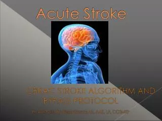
Decision Criteria: The bypass protocol is intended to ensure that patients with a witnessed acute stroke be transported to an accredited stroke center. • Exceptions to the bypass protocol requiring the patient to be transported to the NEAREST facility are: • Inability to establish and/or maintain an airway or in the event of a cardiac arrest. • If transport time to the indicated accredited stroke center exceeds 30 minutes; the patient should be transported to the nearest facility capable of treating stroke with Activase (t-PA) if indicated, then transferred to an accredited stroke facility.
The activation of the Bypass Protocol for the symptomatic acute stroke patient should be initiated upon the recognition of confirmed witnessed changes in patient condition as to “Last Known Well” in less than 4 hours. • If “Last Known Well” temporarily unknown due to patients inability to talk or the lack of a witness, transport to an accredited stroke center and activate a stroke alert.
Hand off of the acute stroke patient to advanced life support “Mobile Intensive Care Unit” or Air Transport will be initiated in the following circumstances: • Basic life support unit is first responder only and/or unable to leave service area • If air transport/pick-up total time is less than ground transport time. HALO Flight (Corpus Christi) 1-800-776-4256 AirLIFE (San Antonio) 1-210-233-5800 PHI (Victoria) 1-877-435-9744 Valley Air (Harlingen) 1-800-679-0911
If >30 minutes by ground to an accredited stroke center or no air medical then transport to the closest facility capable of treating stroke pts. with (t-PA)
Continue airway maintenance and administration of supplemental oxygen. • Obtain IV access if not done prehospital • Central venous catheter • Blood glucose determination • Cardiac monitoring, 12-Lead ECG • Foley catheter
Lab studies • Complete blood count (CBC) with platelet count • Coagulation profile • Serum glucose • Electrolytes, cardiac enzymes • NIH Stroke Scale • Imaging studies • Noncontrast CT of the brain • Differentiates between hemorrhagic and ischemic stroke • Chest X-Ray
Treatment for ischemic stroke may include • Anticoagulants • Antiplatelet agents • Fibrinolytics • Recombinant tissue-type plasminogen activator (rtPA) • Patients with ischemic stroke and hypertension may receive • Labetalol • Enalaprilat • Nicardipine • Nitroglycerin
Treatment of intracerebral hemorrhage • Severe hypertension (MAP >130 mmHg) may be treated. • Labetalol • Enalapril • Nicardipine • Nitroprusside • Increased ICP treated with • Hyperventilation • Mannitol, furosemide • Surgical intervention dependent on patient neurological status plus size and location of hemorrhage
Treatment of subarachnoid hemorrhage • Head elevated to 30 degrees • Maintenance of blood pressure to prehemorrhagic levels • Seizure prophylaxis • Ventriculostomy • Surgical clipping of ruptured aneurysm
Door to Triage by Doctor – 10 minutes • Door to CT Scan – 25 minutes • Door to CT Read/Lab Results – 45 minutes • Door to (t-PA) – 60 minutes