Download
1 / 1
10 likes | 118 Vues
Tx x. Alexander Volovick 1 , Timur Saliev 2 , Sam Eljamel 2 , Roos Eisma 2 , Andreas Melzer 2 1 Insightec , Israel; 2 Institute for Medical Science & Technology, University of Dundee, United Kingdom. Ex-vivo feasibility studies of brain sonication under MR-guidance.

E N D
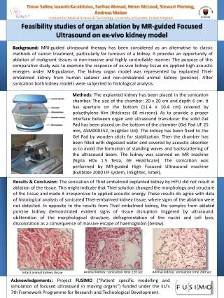
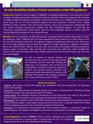
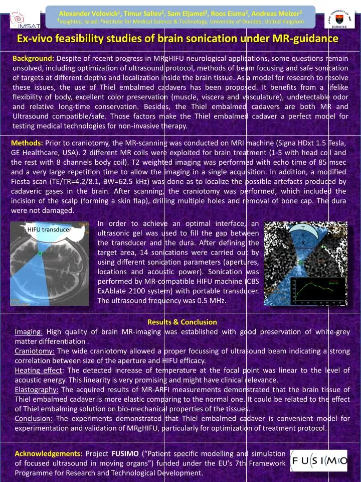
Txx Alexander Volovick1, TimurSaliev2, Sam Eljamel2, Roos Eisma2, Andreas Melzer21Insightec, Israel;2Institute for Medical Science & Technology, University of Dundee, United Kingdom Ex-vivo feasibility studies of brain sonication under MR-guidance Background:Despite of recent progress in MRgHIFU neurological applications, some questions remain unsolved, including optimization of ultrasound protocol, methods of beam focusing and safe sonication of targets at different depths and localization inside the brain tissue. As a model for research to resolve these issues, the use of Thiel embalmed cadavers has been proposed. It benefits from a lifelike flexibility of body, excellent color preservation (muscle, viscera and vasculature), undetectable odor and relative long-time conservation. Besides, the Thiel embalmed cadavers are both MR and Ultrasound compatible/safe. Those factors make the Thiel embalmed cadaver a perfect model for testing medical technologies for non-invasive therapy. Methods: Prior to craniotomy, the MR-scanning was conducted on MRI machine (SignaHDxt 1.5 Tesla, GE Healthcare, USA). 2 different MR coils were exploited for brain treatment (1-5 with head coil and the rest with 8 channels body coil). T2 weighted imaging was performed with echo time of 85 msec and a very large repetition time to allow the imaging in a single acquisition. In addition, a modified Fiesta scan (TE/TR=4.2/8.1, BW=62.5 kHz) was done as to localize the possible artefacts produced by cadaveric gases in the brain. After scanning, the craniotomy was performed, which included the incision of the scalp (forming a skin flap), drilling multiple holes and removal of bone cap. The dura were not damaged. In order to achieve an optimal interface, an ultrasonic gel was used to fill the gap between the transducer and the dura. After defining the target area, 14 sonications were carried out by using different sonication parameters (apertures, locations and acoustic power). Sonication was performed by MR-compatible HIFU machine (CBS ExAblate 2100 system) with portable transducer. The ultrasound frequency was 0.5 MHz. HIFU transducer Results & Conclusion Imaging: High quality of brain MR-imaging was established with good preservation of white-grey matter differentiation . Craniotomy: The wide craniotomy allowed a proper focussing of ultrasound beam indicating a strong correlation between size of the aperture and HIFU efficacy. Heating effect: The detected increase of temperature at the focal point was linear to the level of acoustic energy. This linearity is very promising and might have clinical relevance. Elastography: The acquired results of MR-ARFI measurements demonstrated that the brain tissue of Thiel embalmed cadaver is more elastic comparing to the normal one. It could be related to the effect of Thiel embalming solution on bio-mechanical properties of the tissues. Conclusion: The experiments demonstrated that Thiel embalmed cadaver is convenient model for experimentation and validation of MRgHIFU, particularly for optimization of treatment protocol. Acknowledgements:Project FUSIMO (“Patient specific modelling and simulation of focused ultrasound in moving organs”) funded under the EU's 7th Framework Programme for Research and Technological Development.