Download
1 / 45
650 likes | 1.98k Vues
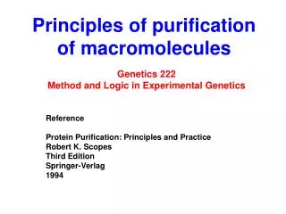
PURIFICATION AND METHODS FOR STUDYING MACROMOLECULES OF LIFE. Aleksander L. Sieroń Department General and molecular biology And genetics http://biolmolgen.slam.katowice.pl. Homogenization (disintegration of cells and tissues). To release: Proteins Nucleic acids.

E N D
PURIFICATION AND METHODS FOR STUDYING MACROMOLECULES OF LIFE Aleksander L. Sieroń Department General and molecular biology And genetics http://biolmolgen.slam.katowice.pl
Homogenization(disintegration of cells and tissues) To release: • Proteins • Nucleic acids
Homogenization(disintegration of cells and tissues) • Homogenization – mechanical procedure • Sonication (disruption of cells by high frequency sound). • Passing the cells through narrow oppening under high pressure. • Permeabilization – making „holes” in cell membrane using gentle detergent. • Pressuring cells using tightly fitted rotating pestle in glass cylinder. • Fragmentation of a tissue using metal balls and high frequency shaking.
SONICATION Homogenization by shaking with metal balls Teflon pestle with tube-like glass cylinders
ISOLATION OF NUCLEIC ACIDS Initial material (biological): • Peripheral blood; • Epithelial cells • Cultured skin fibroblasts • Amniotic fluid cells (AFC), kosmówki (CVS) • Hair follicle cells • Blood spots, semen spots, bone marrow, tissue fragments, skull bone fragments, bone fragments, tooth fragments.
DNA isolation methods • DNA isolation by phenol:chloroform mixture extraction (used for removal of proteins). • DNA isolation with salting out proteins from lysed cells. • DNA isolation using a DNA binding resin to which DNA will be reversibly bound.
RNA isolation methods • Isolation of specific RNA (by total cell RNA fractionation). • Direct isolation of specific RNA (the method is restricted exclusively to the cells synthesizing particular RNA at increased amount). • Poly(A)RNA isolation • Isolation of RNA from polyribosomes. • Total cell RNA isolation with subsequent isolation of particular RNA (cDNA – complementary DNA) by Polymerase Chain Reaction - PCR .
Columns with resin Laboratory micro centrifuge
Quantitative and qualitative analysis After the nucleic acids, DNA or RNA, the firststep is determination of their purity and concentration. Both DNA and RNA concentration can be determined either by UV light absorbance or by comparison of ethidium bromide fluorescence of the sample to control of known concentration
Agarose gel - RNA Agarose gel – DNA after PCR Lanes 1 and 9 contain DNA mass ladder Chromatogram - BIOANALYZER
RNA investigation methods • Northern-blot method • RT-PCR • RT-qPCR • SAGE (serial analysis of gene expression) • Microarrays • Hybridisationin situ (microscopic technique) • RNAseq: High through put transcriptome sequencing
DNA investigation methods Restriction enzymes Hybridization (renaturation): Southern Blotting, DNA fingerprinting, Northern blotting, In situ hybridization (FISH). DNA amplification by PCR. Visualization of PCR products can be achieved by electrophoresis: PCR-Multiplex; PCR-RFLP (analysis of restriction fragments length polymorphism) Sequencing by enzymatic Sanger’s method Parallel sequencing - Next Generation Sequencing
Chromatogram - sequencing Sequencing Polyacrylamide gel
RESTRICTION EcoRI methylase MODYFICATION No cut by EcoRI restriction endonuclease EcoRI restriction endonuclease RESTRICTION
Transfer („blot”) DNA fragments from agarose gel onto membrane NORTHERN BLOT
Isolation steps in proteins purification • The isolation of biological material: • fragmentation of the material, • the crushing of tissues and cells (homogenization, • ultrasound break), • extraction of the aqueous solution of the protein and removal of residual material by centrifugation or filtration • Pre-treatment (optional): precipitation of proteins from solution as a result of fractionation of increasing amounts of solvent or salting out with ammonium sulfate and removing the protein precipitating agent by means of dialysis, centrifugation, differential centrifugation, sedimentation, speed, sedimentation equilibrium • Purification: Chromatography • Changing the ion (optional): dialysis • Analysis of purity of the preparation: electrophoresis, spectrophotometry.
Force of centrifugation Stable sucrose gradient Velocity equilibrium Equilibrium sedimentation Density gradient centrifugation Layers containing particular components can be collected
The four most common types of column chromatography used in protein purification
Examples of selective and non-selective forms of affinity chromatography with the functional group (ligand) used for specificity and typical elution conditions. Summarized from Thermo Fisher Pierce and GE. See below for the common suppliers for different types of columns based on a Labome survey of over 200 publications.
The net charge on a protein is influenced by the pH of its solvent. At pH=pI, the protein has zero net charge and, therefore, will not bind to a cation exchange or an anion exchange stationary phase. Adjusting the pH above or below the pI of the protein will lead to a net charge, and protein binding to either an anion exchange (pH > pI) or a cation exchange (pH < pI) stationary phase.
Sizeexclusioncolumn chromatography used in protein purification
Hydrophobic interaction chromatography. At high ionic strength, proteins are partially desolvated, causing them to adopt alternate conformations in which normally buried hydrophobic residues are more exposed. These residues can then form hydrophobic interactions with the hydrophobic functional groups conjugated to a matrix. Lowering the ionic strength causes the protein to refold into its native conformation, burying its hydrophobic residues. This decreases hydrophobic interactions between the protein and stationary phase, facilitating protein elution
Protein eluted from a hydrophobic interaction column with a decreasing salt (ammonium sulfate) gradient. Fractions were analyzed for both total protein content and activity specific to the protein of interest. The peak centered at fraction 45 contains the protein of interest, as indicated by protein activity. From http://www.insectscience.org/9.04/
Two-demensional polyacrylamide gel electrophoresis Polyacrylamide gel electrophoresis aparatus Polyacrylamide SDS gel electrophoresis – SDS-PAGE
SDS-PAGE of samples collecting during a protein purification scheme. Gel is stained for the visualization of all proteins. From http://www.omicsonline.org/ .
Schematic of a gel for dis-continous gel electrophoresis. Electrolyte composition and pH value of both gels as well as their porosity are different. Concentrating gel Separating gel Buffer – e.g. running buffer Buffer for loading samples - sample buffer Dye front Protein separation according to their isoelectric points
Identification of proteins • Sequencing following: • Fragmentation to shorter fragments (selective protease) • Protein fragment analysis by mass spectrometry
Sample preparation Gel map Dot recovery Pattern analysis e.g. PDQuest Analizadanychprzezporównanie z bazamidanych (Blast, Swissprot) Data analysis by comparison with databases (Blast, Swissprot, etc. Selective protein cutting Protein identification Mass spectrometry
Protein study methods • Crystal formation • Roentgen crystalography X-ray diffraction (analogy to optic microskope) Eye Model of a molecule Computer assisted three-dimension Distribution of electrons: Positions of atoms are reflected by high electron density Crystallographer Computer Phaseinformation Detector X-ray diffraction Crystal Synchrotronic radiation At wave length of X-ray range (0.1 nanometer)
Protein study methods • NMR spectroscopy (Nuclear Magnetic Resonance) Area and PCr line depends on metabolite concentration Line structure is linked to molecule conformation NMR schematic Chemical shift of line Pi depends on pH Sample Magnet Generator Impulse programator Detector Computer Phosphate 31P NMR spectrum from forearm muscle obtained in vivo. Lines in the spectra correspond to nuclei 31P in phospho-ceratine molecules (PCr), inorganic phosphate (Pi) and ATP molecule. Difference of chemical shifts of lines PCr and Pi enables pH measurement.
Backbone model with side chains Ribbon-like model Filed Model
Theoretical Model Solved Structure Three Polish scientists were among the winners of the sixth edition of the prestigous world competition in the modeling of the spatial structure of proteins, which takes place in the United States under the name Critical Assessment of Techniques for Protein Structure Prediction (CASP).
Flowchart of a typical Protein Structure Initiative (PSI) pipeline
CD methods Circular dichroism (CD) Spectroscopy enables: • Determining how the protein is folded (characteristics of secondary, tertiary and even quaternary structure of the protein). • Comparing the structure of proteins obtained from different sources. • Comparing the conformation of the proteins in the various solutions and/or their thermal stability. • Stability study of the conformation of proteins under stress (thermal stability, stability at different pH, in various solvents). • Search for protein conformation changes under the influence of ligands attached.
Comparison of protein structure derived from different sources. Characterization of secondary, tertiary and quaternary protein structures.
Thank you Aleksander L. Sieroń Department General and molecular biology And genetics http://biolmolgen.slam.katowice.pl