Download
1 / 50
540 likes | 879 Vues
PATHOGENESIS AnD research for the PREVENTION OF TYPE 1 DIABETES MELLITUS . NATALIA BOWAKIM ANTA. INDEX. PRESENTATION DIABETES DISEASE DIABETES TYPE 1 STUDIES THAT CAN BE DONE TO PREDICT AND PREVENT DIABETES TYPE 1 BY USING MARKERS : GENETIC IMMUNOLOGIC METABOLIC CONCLUSIONS

E N D
PATHOGENESIS AnDresearch for the PREVENTION OF TYPE 1 DIABETES MELLITUS NATALIA BOWAKIM ANTA
INDEX • PRESENTATION DIABETES DISEASE • DIABETES TYPE 1 • STUDIES THAT CAN BE DONE TO PREDICT AND PREVENT DIABETES TYPE 1 BY USING MARKERS : • GENETIC • IMMUNOLOGIC • METABOLIC CONCLUSIONS • RESEARCH PROTOCOL SETTING TO BE FOLLOWED TO DETECT T1D
INTRODUCTION: • Diabetes mellitus, simply referred to as diabetes—is a group of metabolic diseases in which a person has high blood sugar (hyperglycemia); 2 reasons : • Body does not produce cells do not respond to enough insulin the insulin that is produced • Glycemia regulationis done at islets of Langerhans level [group of endocrines cells located in the pancreas an exocrine gland that secretes the digestive juice in the intestine]
INSULIN • IS : HORMONE THAT ALLOWS THE ENTER OF GLUCOSE IN THE TISSUES. ABSORTION OF Glc • IT’S FORMED BY 2 CHAINS (A and B) joined by 2 sulphuredponts • Its ARNmis traduced in the RE • - formation of its zymogen ; pro-insuline • Transported until Golgi where is intengrated in a clatrinevesicule • Where is fixed, forming insuline and petptice C. • By measuring the quantity of peptide C it’s known the number of alive cells in autoinmune disease.
THE ISLETS OF LANGERHANS (1% of the pancreas) contains different types of cells: • The β cells • in the center of the islet • are metabolic sensors that secretes insulin • represents 60% of the islet • The alpha cells • in the periphery • secretes glucagon (invers effect) • The delta cells • in the periphery • Release somatostatine • The PP cells : • * Releasepolypeptides
ENTRANCE OF GLUCOSE IN β cells • By intermediare transporterGLUT-2 • GlucosephosporilatedbyGLUCOKINASE • Insidetransformed in pyruvateand enter in the Krebs cycleproducingATP • RATIO ATP/ADP Closure of the K-ATP dependantchannel [Can be also done bymedicamentsulphonylurea] • K+ accumulated in theinsideinvolvesdepolarization of themembrane opening of thevoltage Ca2+ channel thefusion of vesiculescontainingtheinsulin release of theinsulineoutsidetheβ cells
Conclusion: If a lot of ATP isproduced [Glucoseblood] high No Glucose receptor
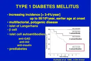
THERE ARE 2 TYPES OF DIABETES TYPE 1 TYPE 2 • 5-10% diabetes • auto-immunedisease • immunesystemkillstheir beta cells receiveinsulineduringtheirlife • usually occurs in childhood and adolescence but can occur at any age • The T-cells recognize the HLA linkage to DQA and DQB genes, and influenced by DRB genes • 80-85% diabetes • No auto-immunedisease • Insulineresistance - hyperglycemia death beta cells. No necessaryinsulineinjection • > 40 years • Geneticpredisposition and diet are theorigin
Causes of beta cellsdisminution TYPE 1 TYPE 2 • PRODUCTION DE IL - 2 BY THE LINFOCITES T HELPER • TRANSCRIPTION OF FACTORS THAT LEAD TO APOPTOSIS - NF-kb and STAT-1 - ER stress JNK, AMP, ROS • DUE TO METABOLIC STRESS [INCREASE OF FATTE ACIDE] DYSFUNCTION IN MITHOCONDRIES DISMINUTION OF REGENERATION BETACELLS
DIABETES TYPE 1 PATHOGENIC
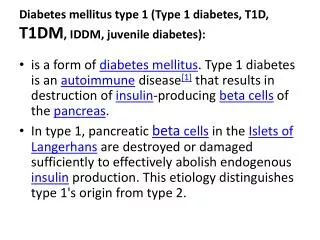
DIABETES TYPE 1insulin-dependent diabetes mellitus (IDDM)andjuvenile diabetes • Concept : autoimmune reaction of the T-cells characterized by destruction of the pancreatic beta cells that leads to the elimination of those one, leading to absolute insulin deficiency • The T-cells recognize the HLA (human leukocyte antigen) which are the major histocompatibility complex in human that are at the surface of the cells
Usually occurs in childhood and adolescence but can occur at any age • Represents 5-10% of the case of diabetes • If this diabetes is not treated by administration of exogenous insulin the child will die in a few weeks • Diabetic ketoacidosis (DKA) can occur in the presence of complete insulin deficiency
The incidence of childhood type 1 diabetes varies based upon : • Geography : highest reported incidences occur in Finland (37 to 45 per 100,000 children younger than the age of 15 years) with rates that are almost 400 times that of Venezuela and parts of China, which have the lowest incidence (0.1 to 0.5 per 100,000 children) • Age: One peak in 4 to 6 years of age and another 10-14 years • Gender: 3:2 male to female ratio even in autoimmune disease are more common in female • Family history: • Monozygotic twin — 50 percent lifetime risk • No familyhistory — 0.4 percent • Offspring of an affected mother — 2 to 4 percent • Offspring of an affected father — 5 to 8 percent • Offspring with both parents affected — reported as high as 30 percent • Environmental factors that increase the risk of developing type 1 diabetes
PREVENTION • No successful strategy for the prevention of type 1 diabetes has yet been identified. • FINALITY OF THE PROJECT : use the COMBINATION OF IMMUNE, GENETIC, AND METABOLIC MARKERS to predict subjects with high risk of develop type 1 diabetes.
BASED ON: ANIMAL MODELS OF TYPE 1 DIABETES • Non-obese diabetic (NOD) mice and BioBreeding(BB) rats are in-bred strains that spontaneously develop autoimmune insulitis and diabetes with similarities to type 1 diabetes in humans. • Several interventions have been tested : • Subcutaneous and oral insulin, nicotinamide, and the ß-cell antigen glutamic acid decarboxylase. • Many interventions have been effective in the murine models when applied before the development of hyperglycemia. • Very few interventions have reversed established diabetes.
BASED ON; PRECLINICAL TYPE 1 DIABETES IN HUMANS A large percentage of the functioning ß-cells must be lost before hyperglycemia appears. Rate of progression : • In some subjects, progression is so slow that diabetes does not occur for many years or perhaps ever. • These subjects regain tolerance: • suppressor T cells become more numerous • helper T cells become less numerous or active. • One report described a 10-year follow-up in 18 nondiabetic twins of type 1 diabetes probands: • the eight twins who developed diabetes had persistently high numbers of CD8 HLA DR+ T-cells, • whereas the 10 twins who remained euglycemic did not
Based on similarities to TD1, in NOD mice : Suppression of insulitis and protection from the development of diabetes can be achieved by: injecting insulin-reactive CD4 T-cell clones from mice that do not develop diabetes into mice that would otherwise have developed diabetes.
BUT THIS VARIABILITY IN PROGRESSION • Causes a therapeutic dilemma with respect to intervention during the preclinical period. • Early therapy • is likely to preserve more ß cells • but some patients being treated unnecessarily. • might increase the risk of type 1 diabetes by disrupting the balance between helper and suppressor activity (a sequence that has been demonstrated in BB rats and NOD mice). • Delaying therapy runs the risk that fewer ß cells will be left to preserve
1. Genetic markers PREDICT TYPE 1 DIABETES
Genetic markers • May be helpful in evaluating the risk of type 1 diabetes in close relatives of a patient with type 1 diabetes. • The risk is markedly increased in these relatives: • about 6% in offspring (children) • 5 % in siblings (brothers and sisters) • 0.4 % in subjects with no family history. • The risk in siblings is importantly influenced by the degree of genetic similarity, • 33 % in identical twins • 12.9 % share two haplotypes • 4.5 % share one haplotypes • 1.8 % no sharing haplotypes
The major susceptibility genes for type 1 diabetes are in the HLA region on chromosome 6p. • Over 90 percent of patients with type 1 diabetes carry • DR4, DQB*0302 and/or DR3, DQB*0201. • Thus, if the proband is heterozygous for DR3 and DR4 (the highest risk combination), the incidence of type 1 diabetes in a sibling who shares these two haplotypes rises to 19 percent. • Diabetes Type 1 unlikely • In the absence of the above alleles makes type 1 diabetes • Especially if the subject carries a protective allele such as DQB*0301, *0602, DRB*0403, or *0406
So, by using the genetic markers plus the family history make possible to estimate the risk of T1D as being as: • low as 1 in 5000 no susceptibility alleles or family history • high as 1in 4 two susceptibility alleles and a positive family history. • The risk for islet autoimmunity drastically increased in DR3/4-DQ2/DQ8 siblings who shared both HLA haplotypes identical
2. IMMUNOLOGIC MARKERS PREDICT TYPE 1 DIABETES Autoantibodies Zinc transporter antibodies
2.1 AUTOANTIBODIES In several prospective family studies in which unaffected first-degree relatives of patients with type 1 diabetes were followed: The presence of ICA [islet-cell antibodies] increased risk of diabetes Particularly if: * ICA titer was high * ICA were persistently detected * ICAwere present in combination with IAA (autoantibodies to insulin)or GAD (antibodies to glutamic acid decarboxylase ):
FINDINGS REPORTED WITH IA-2. [autoantibodies to the tyrosine phosphatases ] • High risk of type 1 diabetes progression for • Those with IA-2 autoantibodies • An autoantibody response directed to the extracellular domain of IA-2 • Suggesting the presence of new antigenic determinants within the extracellular domain of IA-2 . • This has considerable implications : • not only for stratifying high type 1 diabetes risk • but also to facilitate the search for pathogenic epitopes to enable the design of peptide-based immunotherapies, which may prevent the progression to overt type 1 diabetes at its preclinical stages.
Unlike NOD mice, humans exhibit any combination of ICA, IAA, GAD, and IA-2 antibodies. • The risk of type 1 diabetes • is relatively low with IAA alone (Ab to insuline) • is higher with the presence of multiple autoantibodies against islet antigens (insulin, GAD, IA-2 and ICA). • Antibodies to GAD (Abto glutamic acid decarboxylase) are predictive of progression to hyperglycemia even in the absence of ICA or IAA . • As with IAA, the risk is higher in subjects who are ICA-positive
We can conclude that : The risk of diabetes is high in those with multiple autoantibodies (40 % vs3 %in those with single autoantibodies).
2.2 ZINC TRANSPORTER ANTIBODIES • The cation efflux zinc transporter (ZnT8) has also been identified as a candidate type 1 diabetes autoantigen. • 60- 80 % of patients with newly diagnosed type 1 diabetes have ZnT8 autoantibodies. • 26 % of subjects with antibody negative (insulin, GAD, IA-2 and ICA) type 1 diabetes have ZnT8 autoantibodies. • The function of this transporter is unknown. • Alterations in the function of this gene may result in: • abnormal effects on insulin secretion • and/or insulin response.
3.- METABOLIC MARKERS PREVENTION DIABETES TYPE 1
Metabolic markers • Although glucose tolerance remains normal until close to the begginingof hyperglycemia , the acute insulin response to several secretagogues(substance that causes another to be secreted) • Glucose • Arginine • Glucagon • Isoproterenol decreases progressively during the preclinical period. • The most useful and widely performed test is the "first phase" insulin response to glucose (FPIR) during an intravenous glucose tolerance test (IVGTT)
3.1 Intravenous Glucose Tolerance Test (IVGTT) • In this test the rise in serum insulin above baseline is measured during the first 10 minutes after an intravenous glucose challenge. • The response correlates with the functioning ß-cell mass. • The IVGTT • for example, an FPIR below the first percentile of normal: is a strong predictor of type 1 diabetes.
Evaluation of themetabolic factors associated with progression to diabetes • IN THE Diabetes Prevention Trial-Type 1 Diabetes (DPT-1), subjects at high risk for developing diabetes were followed with serial: • IVGTTs (intravenus) • And (OGTTs) oral glucose tolerance tests • Abnormalities of FPIR and two-hour glucose during OGTT had similar sensitivities for diabetes prediction within six months of diagnosis
3.2 Measure the fasting serum concentration of proinsulin • A simpler test that may prove useful the prediction of diabetes is measuring proinsulin the precursor of insulin. • In normal subjects, proinsulin accounts for approximately 15 % of serum immunoreactive insulin. This proportion rises as ß-cell function declines. • In ICA-positive relatives of type 1 diabetes patients serum proinsulin concentrations were three to four times higher compared with ICA-negative relatives
SOLUTIONS TO PREVENT : • Beforethe occurrence of clear signs of islet autoimmunity and type 1 diabetes onset - begging • HLA GENOTYPING AT BIRTH to dentifyindividuals at very high risk of developing type 1 diabetes • RAPID AUTOMATED ASSAYS IN NEWBORNS To screen HLA popultion.
IMMUNONOLOGIC markers • Having tested that : developed diabetes had persistently high numbers of CD8 HLA DR+ T-cells • Whereas subjects that regained tolerance: • Higher number of suppressor T cells • Disminution in number and activity of helper T cells. IN HUMANS Transplatethepancreasisnota solutionto cure TD1 becauseitwillstillkillthe new beta cells
DETECTING clinically useful serum autoantibodies during the preclinical period of type 1 diabetes (Which are the markers of the immune destruction of the β-cell TD1) • islet cell autoantibodies (ICAs) • autoantibodies to insulin (IAAs) • autoantibodies to glutamic acid decarboxylase (GAD6S) • autoantibodies to the tyrosine phosphatases lA-2 and lA-2β • If two or more are positive (Presence of multiple autoantibodies against islet antigens) : • patient should be presumed to have type 1 diabetes . • should be treated with insulin replacement therapy, • as these patients respond poorly to diet and oral hypoglycemic drug therapy. • Particulary the presence of ICA [islet-cell antibodies] (even without affected relatives) in high levels, or in combination with IAA or GAD
Detection of ZnT8 ; • Becauseevenwithoutthe presenceof multiple autoantibodies against islet antigensThe cation efflux zinc transporter (ZnT8) is also a candidate type 1 diabetes autoantigen. • Appearing ZnT8 autoantibodies in the 60- 80 % of patients with newly diagnosed type 1 diabetes. • In the 26 % of subjects with antibody negative (insulin, GAD, IA-2 and ICA)
Using Glucose Tolerance Test as a predictor of T1D • Using an Intravenous Glucose Tolerance Test (IVGTT) an (OGTTs) oral glucose tolerance tests in subjects at high risk for developing diabetes similar sensitivities for diabetes prediction within six months of diagnosis Sensitivity is better when both tests are performed Whereas, Fasting blood glucose levels are a poor predictor of diabetes
ByMeasuring the fasting serum concentration of proinsulin • In normal subjects, proinsulin= 15 % of serum immunoreactive insulin. • In ICA-positive relatives of type 1 diabetes patients serum proinsulinconcentrations = 45–60 %
Researchsettingto be followfor diabetes t1d prevention* protocol
IN A RESEARCH SETTING, THE FOLLOWING APPROACH MAY BE USED : • 1) Test individuals at risk for type 1 diabetes progression for GAD65 and IA-2 autoantibodies. • 2) If they are present and confirmed in a subsequent sample tests for insulin, can be done: • Zinc transporter (ZnT8), • And islet cell antibodies [ICA] • 3) And determine the first phase insulin response to glucose (FPIR).
IN A RESEARCH SETTING, THE FOLLOWING APPROACH MAY BE USED : • 4) The occurrence of multiple antibodies against islet autoantigens serves as a surrogate marker of disease in primary or secondary intervention strategies aimed at halting the disease process. • 5) Genetic typing for susceptibility or protective HLA alleles can also be performed. • This information can be used to determine if a high-risk subject is qualified to be entered into an ongoing prevention trial.