Download
1 / 25
270 likes | 441 Vues
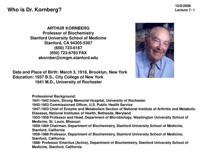
10/9/2006 Lecture 7: 1. Who is Dr. Kornberg?. ARTHUR KORNBERG Professor of Biochemistry Stanford University School of Medicine Stanford, CA 94305-5307 (650) 723-6167 (650) 723-6783 FAX akornber@cmgm.stanford.edu. Date and Place of Birth: March 3, 1918, Brooklyn, New York

E N D
10/9/2006 Lecture 7: 1 Who is Dr. Kornberg? ARTHUR KORNBERG Professor of Biochemistry Stanford University School of Medicine Stanford, CA 94305-5307 (650) 723-6167 (650) 723-6783 FAX akornber@cmgm.stanford.edu Date and Place of Birth: March 3, 1918, Brooklyn, New York Education: 1937 B.S., City College of New York 1941 M.D., University of Rochester Professional Background: 1941-1942 Intern, Strong Memorial Hospital, University of Rochester 1942-1953 Commissioned Officer, U.S. Public Health Service 1947-1953 Chief of Enzyme and Metabolism Section of National Institute of Arthritis and Metabolic Diseases, National Institutes of Health, Bethesda, Maryland 1953-1959 Professor and Head, Department of Microbiology, Washington University School of Medicine, St. Louis, Missouri 1959-1969 Chairman, Department of Biochemistry, Stanford University School of Medicine, Stanford, California 1959-1988 Professor, Department of Biochemistry, Stanford University School of Medicine, Stanford, California 1988- Professor Emeritus (Active), Department of Biochemistry, Stanford University School of Medicine, Stanford, California
10/9/2006 Lecture 7: 2 Dr. Arthur Kornberg and the Department of Biochemistry at Stanford The members of the Stanford Biochemistry Department—Robert Baldwin, Paul Berg, David Hogness, Dale Kaiser, Arthur Kornberg and Robert Lehman—stayed together as a cohesive unit for forty years until retirement. Honors: 1951 Paul-Lewis Award in Enzyme Chemistry 1959 Nobel Prize in Medicine (with Dr. Severo Ochoa) 1965 President, American Society of Biological Chemists 1970 American Philosophical Society 1970 Foreign Member, Royal Society 1979 National Medal of Science 1960 Honorary degrees: City College of New York, Washington University, University of Rochester, Yeshiva University, University of Pennsylvania, University of Notre Dame, Princeton University, Colby College, University of Barcelona, University of Paris, Medical College of Wisconsin, University of Miami 1995 Cosmos Club Award The Nobel Prize in Physiology or Medicine 1959 was awarded for their discovery of the mechanisms in the biological synthesis of ribonucleic acid and deoxyribonucleic acid" Severo Ochoa 1/2 of the prize Arthur Kornberg 1/2 of the prize
10/9/2006 Lecture 7: 3 Dr. Kornberg’s key contributions to nucleic acid research: discovery of DNA polymerase JBC V. 233: 163-170, 1957 We used a 14C labeled substrate of high specific radioactivity and incubated it with ATP and extracts of Escherichia coli, an organism which reproduces itself every 20 minutes. The first positive results represented the conversion of only a very small fraction of the acid-soluble substrate into an acid-insoluble fraction (50 or so counts out of a million added). While this represented only a few ppmoles of reaction, it was something. Through this tiny crack we tried to drive a wedge, and the hammer was enzyme purification.
10/9/2006 Lecture 7: 4 Dr. Kornberg’s key contributions to nucleic acid research: discovery of DNA polymerase Enzyme Assays Assay of "Polymerase"-This assay measures the conversion ~ o of acid-soluble p32-labeled deoxynuc1eoside triphosphates into ~ an acid-insoluble product. The incubation mixture (0.3 mi.) ~ contained 0.02 ml. of glycine buffer (1 M, pH 9.2), 0.02 mI. of a MgCb (0.1 M), 0.03 mi. of 2-mercaptoethanol (0.01 M), 0.02 mi. of~ thymus DNA (0.5 mg. per mI.), 0.01 mi. of dATP (0.5 }Lmole~ per mI.), 0.02 mi. of dGTP (0.5 }Lmole per mI.), 0.01 mi. of'8= dCTP (0.5 J,lmole per mI.), 0.01 ml. of dTP32PP5 (0.5 J,lmole perg mI., 1.5 X 10⁶ c.p.m. per JoImole), and 0.005 to 0.05 unit of to enzyme. Dilutions of the enzyme for assay were made in Tris~ buffer (0.05 M, pH 7.5) containing 0.1 mg. per mi. of thymusg DNA. After incubation at 37° for 30 minutes, the tube was~ placed in ice, and 0.2 mi. of a cold solution of thymus DNA ~ (2.5 mg. per mi.) was added as carrier. The reaction was~ stopped, and the DNA was precipitated by the immediate"N addition of 0.5 mi. of ice-cold 1 N perchloric acid. After 2 to 3 g minutes, the precipitate was broken up thoroughly with a snug¬ fitting glass pestle, 2 mi. of cold distilled water were added, and the precipitate was thoroughly dispersed. After centrifugation for 3 minutes at 10,000 X g, the supernatant fluid was dis¬carded. The precipitate was dissolved in 0.3 ml. of 0.2 N NaOH, the DNA was reprecipitated by the addition of 0.40 mi. of cold 1 N perchloric acid, 2.0 mi. of cold water were added, and the precipitate was thoroughly dispersed. After centrifu¬gation, this precipitate was again dissolved, reprecipitated again and recentrifuged. Finally, the precipitate was dissolved by the addition of 0.2 mi. of 0.1 N NaOH, the entire solution was pipetted into a shallow dish, dried, and the radioactivity meas¬ured.
Dr. Kornberg’s key contributions to nucleic acid research: discovery of DNA polymerase 10/9/2006 Lecture 7: 5 Properties pf E. coli DNA Polymerase I The E. coli DNA polymerase I is a DNA-dependent DNA polymerase that possesses both 3' -> 5' and 5' -> 3' exonuclease activities. It is a single-chain protein with a mass of about 109,000 Da that requires magnesium as a cofactor. Each of its three enzymatic activities are encapsulated into distinct domains of the holoenzyme, such that proteolytic deletions can be generated that lack one or more of the activities. The so-called Klenow fragment is one such molecule that is widely used in recombinant DNA work.
Dr. Kornberg’s key contributions to nucleic acid research: discovery of DNA polymerase 10/9/2006 Lecture 7: 6 Applications of Klenow Fragment 1. Synthesis of double-stranded DNA from single-stranded templates: 2. Filling in recessed 3' ends of DNA fragments: 3. Digesting away protruding 3' overhangs:
Dr. Kornberg’s key contributions to nucleic acid research: discovery of DNA polymerase 10/9/2006 Lecture 7: 7 Applications of Klenow Fragment 4. Preparation of radioactive DNA probes: Examine each of the reactions depicted above. What if the nucleotides used were in the reaction were radioactive? That's correct - the radioactive nucletides would be incorporated into the DNA fragment. Klenow fragment is used frequently to prepare DNA that is labeled with radionuclides or other markers. In some situations, the 3' -> 5' exonuclease activity of Klenow fragment is either undesirable or not necessary. By introducing mutations in the gene that encodes Klenow, forms of the enzyme can be expressed that retain polymerase activity, but lack any exonuclease activity. These forms are the enzyme are usually called exo- Klenow fragment.
Dr. Kornberg’s key contributions to nucleic acid research: Demonstrate the antiparallel orientation of complementary 10/9/2006 Lecture 7: 8 JBC V. 236: 864-875, 1961. ppp*A +pppC+pppG+pppT+DNA template + primers DNA polymerase PPi …pCpTp*ApCpCp*ApGp*ApTpAp*ApTp…. H2O DNase I …+ Cp + Tp* + Ap + Cp + Cp* + Ap + Gp* + Ap + Tp + Ap* + Ap + Tp…. 42 = 16
Dr. Kornberg’s key contributions to nucleic acid research: Demonstration of the antiparallel orientation of complementary strands 10/9/2006 Lecture 7: 9 Predicted relationship of nearest-neighbor dinucleotide frequencies if DNA is antiparallel
Dr. Kornberg’s key contributions to nucleic acid research: Demonstration of the antiparallel orientation of complementary strands 10/9/2006 Lecture 7: 10 Predicted relationship of nearest-neighbor dinucleotide frequencies if DNA is parallel
Dr. Kornberg’s key contributions to nucleic acid research: Demonstration of the antiparallel orientation of complementary strands 10/9/2006 Lecture 7: 11
Dr. Kornberg’s key contributions to nucleic acid research: Demonstration of the antiparallel orientation of complementary strands 10/9/2006 Lecture 7: 12 Radioactivity Radioactive decay produces emitted particles which are capable of ionizing matter. Of particular interest in autoradiography, are beta particles, positrons, and alpha particles: Beta Particles Beta particles show spectra of energies from zero to varying maxima. They produce curved tracks. Positrons Positrons show similar ionizing properties to beta particles. They eventually produce electromagnetic radiation via annihilation. Alpha Particles Alpha particles are strongly ionizing, but lose energy quickly. Since all alpha-emitters are heavy isotopes, they are not used often.
Dr. Kornberg’s key contributions to nucleic acid research: Demonstration of the antiparallel orientation of complementary strands 10/9/2006 Lecture 7: 13 Liquid Scintillation Counting Beta decay, the most common type of radiation used in autoradiography, occurs when a neutron decomposed into a proton and an electron. Positron decay, also of some interest, occurs when a proton converts into a neutron. Some radioisotopes, such as 125I, emit monoenergetic orbital electrons. The theory of beta decay is based on the concepts of neutron or proton conversion, a nuclear process which converts a radioactive substance to one of a more stable atomic state. The first equation below (neutron conversion) is the one most relevant to liquid scintillation.
Dr. Kornberg’s key contributions to nucleic acid research: Demonstration of the antiparallel orientation of complementary strands 10/9/2006 Lecture 7: 14 Liquid Scintillation Counting A common example of this phenomenon is the beta decay of tritium, an element whose nucleus is composed of one proton and two neutrons. One of its neutrons decays into a proton with the ejection of a beta particle and an antineutrino, transforming it into the stable element helium. A similar process occurs within the nucleus of carbon-14, which begins with 6 protons and 8 neutrons but, as a result of beta decay, transforms into stable nitrogen with a nucleus of 7 protons and neutrons each. The total energy of beta decay consists of the energy of the two emitted particles, the beta particle and the antineutrino; this energy is called Emax : Ebeta particle + Eantineutrino = Emax Very few of the emitted beta particles have maximum energy, as energy is shared between the beta particle and the antineutrino. As the diagram below shows, most of the emitted beta particles have an energy of approximately one third of Emax. The maximum energy emitted is actually quite low- for 3H tritium, Emax = 0.0181 MeV, and for 14C, Emax = 0.156 MeV; energy levels which can be easily absorbed by the compound itself, by the surroundings, and by covers on gamma detecting equipment. As a result of the difficulty of detecting the low energy levels generated by beta emission, the technique of liquid scintillation counting developed.
Dr. Kornberg’s key contributions to nucleic acid research: Demonstration of the antiparallel orientation of complementary strands 10/9/2006 Lecture 7: 15 Units of Radioactivity Curie (Ci): the quantity of any radioactive nuclide which undergoes 3.7 x 10¹⁰ disintegrations per second." Units are as follows: A Curie is 1000 millicuries (mCi) A millicurie is 1000 microcuries (µCi) The SI unit of radioactivity is the becquerel (Bq) 1 Bq = 1 disintegration per second = 2.7 x 10⁻¹¹ curies (Ci) 1 Ci = 3.7 x 10¹⁰ Bq = 37 GBq Therefore: 1 mCi = 3.7 x 10⁷ Bq = 37 MBq 1 µCi = 3.7 x 10⁴ Bq = 37 kBq Common Prefixes for SI units 10-3 milli m 103 kilo k 10-6 micro µ 106 mega M 10-9 nano n 109 giga G 10-12 pico p 1012 tera T
Dr. Kornberg’s key contributions to nucleic acid research: Demonstration of the antiparallel orientation of complementary strands 10/9/2006 Lecture 7: 16 The specific activity of labeled compounds refers to the total amount of radioactivity per unit mass, and is commonly quoted in terms of µCi/mg, mCi/mg, Ci/mmol and Bq/mmol. The mass is usually determined by direct weighing or spectroscopic measurements. When there is sufficient mass of a radiolabeled compound for a small sample to be accurately weighed and counted by liquid scintillation counting, the specific activity is expressed as (e.g.) µCi/mg. The conversion from µCi/mg to mCi/mmol is by multiplying the molecular weight and dividing by 1000. When the specific activity is greater than 1 Ci/mmol there is often insufficient material present to be weighed. If the specific activity is greater than 1 Ci/mmol, the specific activity may be calculated by relating the radioactive concentration (determined by liquid scintillation counting) to the chemical concentration, and then converting the figure obtained to Ci/mmol (Bq/mmol). The chemical concentration is commonly determined by U.V. spectroscopy or an appropriate colorimetric method. Specific Activity(mCi/mmol) = total activity (mCi) x molecular weight (mg/mmol) / mass (mg) [α-32P]dATP (3000 Ci/mmol) from Amersham International, 250 µCi or 1 mCi [α-32P]dATP (800 Ci/mmol) from ICN, 250 µCi or 1 mCi
Dr. Kornberg’s key contributions to nucleic acid research: Demonstration of the antiparallel orientation of complementary strands 10/9/2006 Lecture 7: 17 Radioactive isotopes frequently used in biomedical sciences include: 32P; a beta-emitter (1.71 MeV) with a half-life of 14.3 days which is used routinely in life-science laboratories, primarily to produce radiolabeled DNA and RNA probes, e.g. for use in Northern blots or Southern blots. Because the high energy beta particles produced penetrate skin and corneas, and because any 32P ingested, inhaled, or absorbed is readily incorporated into bone and nucleic acids, OSHA requires that a lab coat, disposable gloves, and safety glasses or goggles be worn when working with 32P, and that working directly over an open container be avoided in order to protect the eyes. Monitoring personal, clothing, and surface contamination is also required. In addition, due to the high energy of the beta particles, shielding this radiation with the normally used dense materials (e.g. lead), gives rise to secondary emission of X-rays via a process known as Bremsstrahlung, meaning braking radiation. Therefore shielding must be accomplished with low density materials, e.g. Plexiglas, acrylic, Lucite, plastic, wood, or water. 33P; a beta-emitter (0.25 MeV) with a half-life of 25.4 days. It is used in life-science laboratories in applications in which lower energy beta emissions are advantageous such as DNA sequencing. 14C and 3H are the two most commonly used nuclides The choice between 14C and 3H is complex 3H gives better resolution due to low ß energies 3H has a higher specific activity, minimizing the necessary dose 3H's half-life is 500 times shorter than that of 14C (5800 years) Tritiated substances are 500 times as hot as 14C labeled substances However, tritiated substances are often less reliable Until recently, 14C has dominated in whole-body autoradiography due to higher efficiency Radioactive 3H doses of 20 times that of 14C give about the same exposure time Since tritiated substances are 10 to 20 times less expensive, the cost per dose is about the same 125I is also commonly used in both gross and microautoradiography 125I emits extranuclear electrons of discrete energies Earlier, 35S had been commonly used 35S has properties similar to 14C, except that it has a short half-life of 87 days
Dr. Kornberg’s key contributions to nucleic acid research: Demonstration of the antiparallel orientation of complementary strands 10/9/2006 Lecture 7: 18 Liquid Scintillation Counting Used for the measurement of beta emitting nuclides, such as, tritium, carbon-14, and phosphorus-32. This technique involves dissolving the sample containing a radionuclide in a suitable scintillation solution and the use of a liquid scintillation counter. The solution normally consists of an aromatic organic solvent (historically benzene or toluene, but more recently less hazardous solvents have come into favour) containing a fluor and a detergent to make the whole solution miscible when counting aqueous samples. The energy of the emitted beta particles is transferred via the solvent to the primary fluor and sometimes to a secondary fluor, which then emits energy as light photons. These photons are detected using a photomultiplier. Only a small proportion of the available energy is liberated as light. The residue is dissipated as vibrational and rotational energy in the solvent. S4023, Sigma Sigma-Fluor™ High Performance LSC Cocktail, For aqueous samples High performance cocktail with a high flash point (120 °F / 49°C) and lower vapor pressure than xylene or toluene based cocktails for improved safety in use and storage. Counting efficiencies under ideal conditions range from about 30% for Tritium (a low-energy beta emitter) to nearly 100% for Phosphorus-32, a high-energy beta emitter. Some chemical compounds (notably Chlorine compounds) and highly colored samples can interfere with the counting process. This interference, known as "quenching", can be overcome through data correction or through careful sample preparation.
Dr. Kornberg’s key contributions to nucleic acid research: Demonstration of the antiparallel orientation of complementary strands 10/9/2006 Lecture 7: 19 High-energy beta emitters such as P-32 can also be counted in a scintillation counter without the cocktail. This technique, known as Cherenkov counting, relies on the Cherenkov radiation being detected directly by the photomultiplier tubes. Cherenkov counting in this experimental context is normally used for quick rough measurements, since it is more liable to variation caused by the geometry of the sample. Cherenkov radiation (also spelled Cerenkov or sometimes Čerenkov) is electromagnetic radiation emitted when a charged particle passes through an insulator at a speed greater than the speed of light in that medium. The characteristic "blue glow" of nuclear reactors is due to Cherenkov radiation. It is named after Russian scientist Pavel Alekseyevich Cherenkov, the 1958 Nobel Prize winner who was the first to rigorously characterize it.
Dr. Kornberg’s key contributions to nucleic acid research: Demonstration of the antiparallel orientation of complementary strands 10/9/2006 Lecture 7: 20 Benzene has been chosen because of its excellent light transmission properties and the high chemical conversion yield of sample C to benzene. Synthesis of Benzene includes: The carbon is first oxidised to CO2, either by acid hydrolysis (for carbonates), or combustion in an oxygen stream or combustion bomb (for organic materials). 2CO2 + 10Li => Li2C2 + 4Li2O Li2C2 + 2H2O => C2H2 + 2LiOH 3C2H2 => C6H6 Commercially available LS vials used for benzene counting are commonly composed of either Teflon, quartz or low-K glass. Polyethylene and polypropylene vials are very useful for non-aromatic solvents, but their permeability makes them unsuitable for repeatable long-term benzene use.
Dr. Kornberg’s key contributions to nucleic acid research: Demonstration of the antiparallel orientation of complementary strands 10/9/2006 Lecture 7: 21 Liquid Scintillation Counting The photoelectric effect is the emission of electrons from matter upon the absorption of electromagnetic radiation, such as ultraviolet radiation or x-rays. An older term for the photoelectric effect was the Hertz effect, though this phrase has fallen out of current use. Einstein: light quanta Albert Einstein's mathematical description in 1905 of how it was caused by absorption of what were later called photons, or quanta of light, in the interaction of light with the electrons in the substance, was contained in the paper named "On a Heuristic Viewpoint Concerning the Production and Transformation of Light". This paper proposed the simple description of "light quanta" (later called "photons") and showed how they could be used to explain such phenomena as the photoelectric effect. The simple explanation by Einstein in terms of absorption of single quanta of light explained the features of the phenomenon and helped explain the characteristic frequency. Einstein's explanation of the photoelectric effect won him the Nobel Prize (in Physics) of 1921. quantum mechanics In a more general sense: synonymous with quantum theory. In a more restricted sense: The quantum theory of particles moving under the influence of forces - wherein the particles are described as quantum objects, while the forces are not. An important application of quantum mechanics is the physics of the electron shells of atoms ("atomic physics", for short). Attempts to extend the quantum laws to govern the forces themselves lead to relativistic quantum field theories.
Dr. Kornberg’s key contributions to nucleic acid research: Demonstration of the antiparallel orientation of complementary strands 10/9/2006 Lecture 7: 22 Liquid Scintillation Counting Photomultiplier A device to convert light into an electric signal (the name is often abbreviated to PM). Photomultipliers are of great relevance in all detectors based on scintillating material. A photomultiplier consists of a photocathode (photons are converted into electrons, making use of the photoelectric effect), a multiplier chain (strings of successive electron absorbers with enhanced secondary emission, called dynodes , the entire string using electric fields to accelerate electrons), and an anode, which collects the resulting current. A dynode is one of a series of electrodes within a photomultiplier tube. Each dynode is more positively charged than its predecessor. Secondary emission occurs at the surface of each dynode. Such an arrangement is able to amplify the tiny current emitted by the photocathode by typically one million.
Dr. Kornberg’s key contributions to nucleic acid research: Demonstration of the antiparallel orientation of complementary strands 10/9/2006 Lecture 7: 23 PMTs are similar to phototubes. They consist of a photocathode and a series of dynodes in an evacuated glass enclosure. Photons that strikes the photoemissive cathode emits electrons due to the photoelectric effect. Instead of collecting these few electrons (there should not be a lot, since the primarily use for PMT is for verly low signal) at an anode like in the phototubes, the electrons are accelerated towards a series of additional electrodes called dynodes. These electrodes are each maintained at a more positive potential. Additional electrons are generated at each dynode. This cascading effect creates 105 to 107 electrons for each photon hitting the first cathode depending on the number of dynodes and the accelerating voltage. This amplified signal is finally collected at the anode where it can be measured.
Dr. Kornberg’s key contributions to nucleic acid research: Demonstration of the antiparallel orientation of complementary strands 10/9/2006 Lecture 7: 24 Liquid Scintillation Counting
Dr. Kornberg’s key contributions to nucleic acid research: Demonstration of the antiparallel orientation of complementary strands 10/9/2006 Lecture 7: 25 Geiger Counter An ion or electron penetrating the tube (or an electron knocked out of the wall by X-rays or gamma rays) tears electrons off atoms in the gas, and because of the high positive voltage of the central wire, those electrons are then attracted to it. In doing so they gain energy, collide with atoms and release more electrons, until the process snowballs into an "avalanche" which produces an easily detectable pulse of current. With a suitable filling gas, the flow of electricity stops by itself, or else the electrical circuitry can help stop it. The instrument was called a "counter" because every particle passing it produced an identical pulse, allowing particles to be counted (usually electronically) but not telling anything about their identity or energy (except that they must have sufficient energy to penetrate the walls of the counter).