Protein
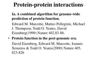
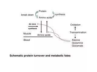
Protein. synthesis. break down. Amino acids. de novo synthesis. Oxidation. Transamination. Muscle. Amino acids. Blood. Alanine Glutamine Glutamate. Schematic protein turnover and metabolic fates. Muscle will not improve with protein feeding alone!.

Protein
E N D
Presentation Transcript
Protein synthesis break down Amino acids de novo synthesis Oxidation Transamination Muscle Amino acids Blood Alanine Glutamine Glutamate Schematic protein turnover and metabolic fates
Muscle will not improve with protein feeding alone! Fed-state gains and fasted state losses in muscle protein balance Skeletal muscle mass is maintained by normal protein feeding. Feeding refreshes muscle protein to improve muscle function, permitting more physical activity. Overfeeding of protein increases insulin resistance- muscle proteolysis Fed state gains are enhanced, fasted state losses are less Improvement in immune function stimulation of protein synthesis
Degradation of amino acids rich protein diet: Catabolised for energy, postprandial gluconeogenesis stored as liver glycogen biosynthetic reactions Glucose-6P-ase PEPCK excreted directly in the urine AMP purine nucleotide cycle Starvation, catabolic states: Gluconeogenesis PEPCK Glucose-6P-ase IMP (liver) blood kidney urine
Central role of glutamate in nitrogen metabolism Ser Thr His + amino acid a-keto-glutarate NH4 oxidative deamination transamination Asp Glutamate keto acid Gln • Amino group from the majority of amino acids is collected by glutamate (by transamination) • in the hepatocytes. • - Liberation of the amino group in the formation of NH+4 by GDH.
L-glutamate dehydrogenase reaction Glutamate in amino acid synthesis, degradation and interconversion glutamate glutamine other amino acids NH4+ intestinal bacteria Allosteric regulation of glutamate dehydrogenase
Catabolism of L-amino acids Transaminases (aminotransferases) GOT Glutamate + oxaloacetate (OAA) <---> a-ketoglutarate + aspartate ASAT GPT Glutamate + pyruvate <---> a-ketoglutarate + alanin ALAT Coupled transamination reaction
PRP Pyridoxal phosphate (PRP) and PRP in aldimine linkage to the lysine residue of the transaminase (Schiff-base)
Different forms of pyridoxal phosphate during a transamination reaction R1- R2 E R2- R1 pyridoxamine phosphate
Specific pathways for the deamination of amino acids (minor routes) Serine dehydratase Metabolism of serine for gluconeogenesis cystein desulphhydrase D-amino oxidases (FAD), L-amino acid oxidase (FMN)
Transport of ammonia Concentration of ammonia in the systemic blood is very low (25-50μmol/L), toxic to the brain. Transport: glutamine and alanine (muscle) glutamine ( brain) Glutamine: non-toxic carrier 0.5-0.8mM in arterial plasma, 20-25% of circulating free amino acids precursor for synthesis of many nitrogen containing compounds metabolic fuel for rapidly dividing cells generates glutamate and GABA in the brain
Glutamine transport , interorgan metabolism of glutamine de novo synthesis: L-Glutamate + NH4+ + ATP L-Glutamine + ADP+ Pi compartmentalised glutaminase glutamine synthetase Glutamine in diet low glutaminase Glutamine - principle non-toxic carrier of nitrogen Intracellularly – muscle pool – released in response to stress, hypercatabolic states brain – glutamine-glutamate cycle- GABA liver – catabolised - substrate for ureagenesis and gluconeogenesis kidney – catabolised - ammoniagenesis and gluconeogenesis muscle, lung, adipose - major sites of glutamine release to blood
glutamine synthetase Muscle Lung/adipose Muscle release Plasma Glutamin uptake Liver Glutamin proline, ornithine,citrulline, alanine Gut carbon sceleton: glycogen glucose NH+4 urea Brain Kidney Glutamate- glutamine cycle acid-base balance NH3 portal vein liver glutamine+H2O glutamate + NH 3 glutaminase
The liver receives both amino acids and ammonia from circulation Scource of ammonia in different tissues: 1. degradation of amino acids transdeamination (transamination+GDH) minor patways 2. deamination of other compounds N-containing side chains of nucleotides neurotransmitters 3. ammonia production in the large intestine by bacteria portal vein, direct transport of ammonia. Urea cycle Function: 1. prevents ammonia levels from rising too high when large amounts of amino acids are catabolized 2. urea cycle enzymes: extrahepatic arginine synthesis
Biosynthesis of urea in the liver 55-100g protein/day ORNT1 ORNT1
The liver receive both ammonia and amino acids from the circulation GDH and major aminotransferases catalyze reactions close to equilibrium Quantitative aspects of nitrogen incorporation, regulation?
Regulation of the urea cycle 1. Short term: NAG an allosteric regulator of CPSI and glutaminase activity increased amino acid catabolism increase in NAG + glutaminase increased flux with constant ammonia concentration increase in glutamate, more NAG + Arginine mitochondria 2. Long term: high protein diet: transcriptional regulation. Hepatic glycogen syntesis Caloric restriction: increased protein catabolism – CPSI induction (cAMP responsive element), glucose need. ORNT1 - increased transcription.
Hyperammonemias deffect: carbamoyl phosphate synthetase CPSD CP cytosol, pyrimidine synthesis, orotic acid deffect: ornithine transcarbamoylase OTCD NH4 +
Inherited urea cycle diseases (+liver failure) Having no urea cycle, brain relies on glutamine synthetase for the removal of exes ammonia Hyperammonemia Brain edema, convulsions, coma NH3 Change in astrocyte morphology: cell swelling astrocytosis acute hyperammonemia chronic hyperammonemia Changes in expression of glutamate transporters in astrocytes.
Scriver et.al.The metabolic and Molecular Bases of Inherited Deseases,2001
Hyperammoniemic encephalopathy Brusilov: Rev. in Mol. Medicine,2002 Computer axial tomography scan of the head of hyperammonemic encephalopathy in the composite case of ornythine transcarbamoylase deficiency. A. CT within normal limits upon admission B. CT scan after tonic seizure with bilateral hemispheric edema with effacement of cerebrospinal fluid spaces.
The actrocyte demonstrating its relationship with other structures in the brain Brusilov: Rev.in Mol. Medicine,2002
The glutamate synapse, effect of NH3 on the the Glutamate-glutamine cycle intracellular Glu depletion extracellular accumulation glutaminase glutamine synthetase NH3 Ca2+ NO Brain injury Felipo et.al.:Progress in Neurobiology,2002
Treatment: - limited nitrogen diet - arginine becomes an essential amino acid - detoxification reactions as alternatives to the urea cycle, ATP dependent
Hepatic metabolism of glutamine, zonal distribution of glutaminase and glutamine synthetase bulk remaining detoxify glutaminase glutamine synthetase high affinity spare aminonitrogen in starvation high capacity low affinity at metabolic acidosis: net producer of glutamine Sequential synthesis of urea and glutamine – efficient to ensure systemic/nontoxic level of ammonia Ammonium ion - feed-forward activator of synthesis of glutamate and N-acetyl glutamate Hepatic synthesis of glutamine – acid-base balance. Decrease pH – activation of glutamine synthetase –sparing of glutamine
Interorgan metabolism of glutamine during metabolic acidosis Acut response: plasma glutamine Renal extraction of glutamine glutamine synthetase release incresased ammonia excretion uptake increased gluconeogenesis PEPCK pH
The urea cycle – part of the metabolism centered around L-arginine
Bioavailability of arginine is complex 1.Exogenous supply 2.Endogenous release 3.Arginine resynthesis 4.Arginine catabolism, arginase 5.Arginine transport L-arginine is semiessential amino acid, synthesized in collaboration. The intestinal – renal axis. Arg CAT-1 AS, AL urea arginase EC, nerve cells, macrophages Arg CAT-1 circulation Arg NO+citrulline Insufficient Arg: strict carnivors small bowel, kidney disease conditions with elevated amino acid catabolism: inflamation, sepsis, recovery.
Arginine availability: arginases and NOS use a common substrate Fate of citrullin: intercellular citrulline-NO cycle - cell proliferation repair Citrulline recycled to Arg, in kidney +other tissues inflammatory stimuli
Arginine is the largest scource for NO production NO,(EDRF): labile, common gas NO-cGMP-mediated effects: smooth muscle cell relaxation in EC: cGMP-prostacyclin mediated decrease in platelet aggregation decrease in leukocyte adhesion and migration NO functionality – vascular health/vasculopathy - production of NO – depends on NOS activity “Arginine paradox” Km for eNOS: 1.4-2.9 μmol/L Intracellular L-arginine: 0.5-2mmmol/L eNOS should be saturated with substrate Despite high cellular arginine, and low Km of eNOS: arginine, citrulline supplementation “in vivo” improves NO function: increased vasodilation decreased leukocyte adhesion decreased platelet adhesion Possible reasons: altered arginine transport increased arginase activity compartmentalisation of arginine
Supplementation: Arg: low bioavailability, increased arginase Cit: Arg synthesis, increased NO levels Gln: major vehicle of transport, Glu-gluthatione reduction of oxidative stress Gly: restores NO balance at increased nutrient demands Meth, Homocys: increased cardiovascular risk Lys: decreases Arg transport