Download
1 / 38
420 likes | 784 Vues
Acquired oculomotor nerve palsy in childhood - SCHWANNOMA. Dr. Divyaswathi Citla Sridhar, PGY-1, Pediatrics Dr. Elisa Rhee , PGY-1, Pediatrics Pediatric Neurology Grand rounds 07.25.2014. Chief complaint Left eye ptosis, blurry vision and eye pain History of presenting illness

E N D
Acquired oculomotor nerve palsy in childhood - SCHWANNOMA Dr. Divyaswathi Citla Sridhar, PGY-1, Pediatrics Dr. Elisa Rhee , PGY-1, Pediatrics Pediatric Neurology Grand rounds07.25.2014
Chief complaint • Left eye ptosis, blurry vision and eye pain • History of presenting illness • 7 y/o right handed African American girl • Had left eye pain for the past 3 days, woke up at around 10am on 7/1/14 with “left droopy eyelid” with blurry vision. • Reports double vision on lateral gaze bilaterally that corrects when either eye is covered. • Per dad, similar event at 6 months of age (left ptosis) that corrected by itself.
Review of symptoms • GEN: + fatigue, + low grade fever • EYES: + blurred vision, + double vision, + eye pain in left eye • PULM: + cough • GI: + vomiting • NEURO: As per HPI
PMH/PSH • Had cough 1 week back, possible pneumonia treated with oral antibiotics. • Sickle cell anemia (parents have sickle cell trait) • Meds: Cetirizine, Iron pills, Antibiotic (unknown) • Family history: Mother and father with sickle cell trait
Physical Examination • Vitals: Stable, no fever • Ht: 89.8%; Wt: 25% • Awake, alert, active • HEAD: Normocephalic and atraumatic
Left eye 1. Ptosis 2. WEAKNESS on • Elevation (superior rectus), • elevation/adduction (inferior oblique), • adduction (medial rectus), • depression/adduction (superior oblique) 3. Pupils: L: 5mm -> 3mm R : 4mm -> 2mm
Rest of the physical exam – normal • NEURO Cranial nerves: II- XII intact except 3rd nerve Motor: normal Sensory: intact Gait: normal Cerebellum: normal
Differential Diagnosis • Infection: meningitis/ encephalitis/ brain abscess • Mass lesion compressing CN III • Vascular process: midbrain stroke (secondary to sickle cell disease) • Aneurysm:PCA/ basilar • Migraine: ophthalmoplegic • Auto-immune disease: SLE, myasthenia gravis • Demyelineating disease: post viral • Trauma
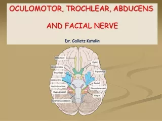
CN III; Somatic Motor (oculomotor output) Also… (let’s not forget) Levator palpebrae superioris; 2 innervations
Anatomy of CN III – cross section Parasympathetic output (Visceral motor; superiomedial) • Oculomotor output • (Somatic motor) Vascular lesions Compression (e.g. SOL)
Etiology of CN III palsy • Nuclear lesions • Lesion in one SR subnucleus Bilateral • Lesion on nerve SR fascicles Unilateral • Lesion on central caudal site Ptosis • Lesion on rostral site Mydriasis (pupil) • Lesion on internuclear neurons Contralateral abduction weakness • Etiologies: congenital hypoplasia, infarction/hemorrhage, tumor, trauma, infection
Congenital CN III palsy • Incomplete and unilateral; aberrant reinnervation • Rarely, CN III palsy with cyclic spasms • Variable lesion locations • Ophthalmoplegic migraine: • Childhood onset • Complete CN III palsy, lasts days or weeks • Schwannomaof CN III: • Slowly progressive • Cerebrovascular malformation: • Infants
Etiology of CN III palsy • Lesions on Fascicles • Lesion on nerve fascicles Unilateral • Etiologies: Infarction, hemorrhage, demyelination, syphilis, trauma • Lesions on Subarachnoid portions • Aneurysm (PCommA) Usually pupillary defect Cf. nerve infarction (does not need arteriography) • Lesions at the tentorial edge • Transtentorial herniation Pupillary defect(mydriasis)
Etiology of CN III palsy • Lesions on the cavernous portion • Aneuryms progressiveophthalmoplegia and ptosis (with signs of aberrant reinnervation); 50% facial pain • Tumors (meningioma, pituitary adenomas, lymphomas) without any pain! • Hemorrhage: pituitary apoplexy • Infarction of the CN III “medical third” • Sparing/minimal involvement of pupil • Facial/orbital pain: • Followed by muscle palsy (up to 2 weeks to evolve) • Disappear when diplopia or ptosis develops (sudden) • Recovery within 3 months • A/w diabetes, HTN, collagen vascular disease, giant cell arteritis
Etiology of CN III palsy • Partial CN III palsy • Isolated lesions of the terminal branches • Note. Isolated ptosis or paralysis of individual muscles – Consider myasthenia gravis
Our Patient – Diagnostic tests • MRI Brain – No obvious radiological abnormality • MRA Head and Neck – Normal • MRI Orbit (w/wo contrast): • At the left interpeduncular fossa between the left P1 segment and superior cerebellar artery there is an enhancing 4 mm focal, round lesion involving the proximal left oculomotor nerve favored to represent a schwannoma. No connection to the vasculature seen.
Coronal; T1 pre-contrast Coronal; T2 pre-contrast
Other tests & course performed • Lumbar puncture • CSF studies not remarkable (colorless, clear, G-55/P-24/WBC-1/RBC-0) • Ruled out, treatable infection/inflammatory etiology • Started, 5 day course of steroids • IV Methlyprednisolone 750mg/day (30mg/kg/day) • MRI spine • No mass lesion; epidural lipomatosis in the upper to mid-thoracic spine with mild compression of the thecal sac • Ruled out schwannoma at other sites (neurfibromatosis)
The patient was discharged with… • 10 day tapering steroid • 50mg/day x 2d 40mg/day x 2d 30mg/day x 2d 20mg/day x 2d 10mg/day x 2 d stop • Follow up with Dr. John Slopis at MDA (director of NF clinic) • Follow up with ophthalmology with Dr. Kumar
Case Studies • 41 well-documented individuals with isolated oculomotor nerve schwannoma in the literature • Of these only 13 cases occurred in children under the age of 18 [1-13].
A 3 year old boy with ptosis in the right eye for 2 weeks. • MRI: 11*12*13 mm right parasellar and suprasellar mass with homogenous enhancement, which medially compressed the right uncusslightly without displacing the pituitary stalk and the brainstem
Classification Watch for increase in size during follow up
Management • Total tumor resection – 9 cases out of 13 8 – Aggravating oculomotor nerve dysfunction postoperatively 1 – Improving nerve function (intra orbital tumor) • Indication for surgical treatment: Large tumors that presented in association with consciousness disturbance, other cranial nerve signs, hemiparesis due to mass effect, malignant features with rapid enlargement. • Oculomotor nerve grafting after tumor resection: Inadequate recovery of function due to imprecise functional distribution of the grafted nerve.
To increase the chance of functional preservation of the oculomotor nerve • Partial Resection & Follow up: can resect tumor within the oculomotor cistern follow-up for the more anterior residual part of the tumor • Gamma Knife (Elekta Corp, Stockholm, Sweden) and proton beam therapy treatment for the remaining tumor or microneuroma Radiobiological mechanism: combination of direct tumoricidal effects and intratumoral vascular obliteration • A wait-and-see policy is recommended in asymptomatic cases or microneuroma (15)
References: 1. Chewning RH, Sasson AD, Jordan LC, Tamargo RJ, Gailloud P. Acute third cranial nerve palsy from a third cranial nerve schwannoma presenting as a saccular aneurysm on three-dimensional computed tomography angiography: Case illustration. J Neurosurg 2008;108: 1037. 2. Leunda G, Vaquero J, Cabezudo J, Garcia-Uria J, Bravo G. Schwannoma of the oculomotor nerves. Report of four cases. J Neurosurg 1982;57:563e5. 3. Muhammad SS, Muhammad Ei, Khalid NC, Asad A. A child with intra-orbital oculomotor nerve schwannoma without neurofibromatosis. Can J Neurol Sci 2008;35:528e30. 4. Bejjani GK, Duong DH, Kalamarides M, Ziyal I, Sullivan BJ. Cerebral vasospasm after tumour resection. A case report. Neurochirurgie 1997;43:164e8. 5. Bisdorff AR, Wildanger G. Oculomotor nerve schwannoma mimicking ophthalmoplegic migraine. Cephalalgia 2006;26: 1157e9 6. Kansu T, Ozcan OE, Ozdirim E, Onol B, Gurcay O. Neurinoma of the oculomotor nerve. Case report. J Clin Neuro Ophthalmol 1982;2: 271e7. 7. Kozic D, Nagulic M, Ostojic J, et al. Malignant peripheral nerve sheath tumor of the oculomotor nerve. Acta Radiol 2006;47:595e8. 8. Mariniello G, Horvat A, Dolenc VV. En bloc resection of an intracavernous oculomotor nerve schwannoma and grafting of the oculomotor nerve with sural nerve. Case report and review of the literature. J Neurosurg 1999;91:1045e9 9. Murakami T, Funatsuka M, Komine M, et al. Oculomotor nerve schwannoma mimicking ophthalmoplegic migraine. Neuropediatrics 2005;36:395e8. 10. Netuka D, Benes V. Oculomotor nerve schwannoma. Br J Neurosurg 2003;17:168e73
11. Niazi W, Boggan JE. Schwannoma of extraocular nerves: survey of literature and case report of an isolated third nerve schwannoma. Skull Base Surg 1994;4:219e26. 12. Sener RN. Malignant oculomotor schwannoma: Diffusion MR imaging. J Neuroradiol 2006;33:270e2. 13. Carlow TJ. Oculomotor ophthalmoplegic migraine: Is it really migraine? J Neuro-Ophthalmol 2002;22:215e21. 14. Celli P, Ferrante L, Acqui M, Mastronardi L, Fortuna A, Palma L. Neurinoma of the third, fourth, and sixth cranial nerves: A survey and report of a new fourth nerve case. Surg Neurol 1992;38: 216e24. 15. Asaoka K, Sawamura Y, Murai H, Satoh M. Schwannoma of the oculomotor nerve: A case report with consideration of the surgical treatment. Neurosurgery 1999;45:630e3. 16. Yang SS1, Li ZJ, Liu X, Li Y, Li SF, Zhang HD. Pediatric Isolated Oculomotor Nerve Schwannoma: A New Case Report and Literature Review; Pediatr Neurol. 2013 Apr;48(4):321-17. 17.http://www.bmc.med.utoronto.ca/cranialnerves/?page_id=22 18.http://www.britannica.com/EBchecked/topic/528563/Theodor-Schwann
Theodor Schwann (7 December 1810 – 11 January 1882) was a German physiologist • considered “Founder of modern histology” • Contributions : • A cofounder (with Matthias Schleiden) of the cell theory • Discovery of Schwann cells in the peripheral nervous system • Discovery and study of pepsin, the first digestive enzyme prepared from animal tissue • Discovery of the organic nature of yeast • Invention of the term metabolism.