Chromosomal Abnormalities
Chromosomal Abnormalities. Dr. Attya Bhatti. Chromosomal Abnormalities. Any change in the normal structure or number of chromosomes; often results in physical or mental . Chromosome Abnormalities. Basic categories : Chromosome rearrangements :


Chromosomal Abnormalities
E N D
Presentation Transcript
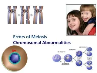
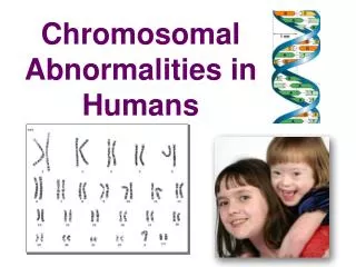
Chromosomal Abnormalities Dr. Attya Bhatti
Chromosomal Abnormalities • Any change in the normal structure or number of chromosomes; often results in physical or mental
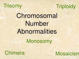
Chromosome Abnormalities • Basic categories : • Chromosome rearrangements: • Chromosome rearrangements alter the structure of chromosomes; for example, a piece of a chromosome might be duplicated, deleted, or inverted. • Aneuploids: • the number of chromosomes is altered: one or more individual chromosomes are added or deleted. • Polyploids: • one or more complete sets of chromosomes are added.
HETEROPLOIDY ABERRATIONS • Chromosomal aberrations that changes the ploidy (one set of chromosomes) of an organisms is called heteroploidy. • Euploid Aberrations • Aneuploid Aberrations
Numerical Abnormalities • Involves the loss or gain of one or more chromosomes, referred as Aneuploidy • Euploid/ Polyploidy
EUPLOID ABERRATIONS • Euploid mutations produces organisms possessing multiple sets of chromosomes. • These are the changes in the number of chromosomes.
Monoploids • One set of chromosomes (n=ABC) is present, mostly, in the nuclei of haplotonic organisms. • E.g. Chlamydomonas • Neurosopora • Also in diploid organisms, is usually in sex cells (male bees and wasps)
Triploids • Organisms may receives three sets of chromosomes(3n= AAA-BBB-CCC). • Results due to union of haploid gamete with diploid gametes. • These organisms are sterile and not common in Natural populations.
Tetraploids • Tetraploid organisms have four sets of chromosomes(4n= AAAA-BBBB-CCCC). • Arises in body cells by the somatic doubling of chromosomes number. • Produced by the union of diploid gametes.
Two groups • Autotetraploids (auto-self): Produced by either somatic doubling of homologous chromosomes, or by the union of diploid gametes of the same species. • Parental genotype AABBCC X AABBCC AAAABBBBCCCC
Allotetraploids (Allo: non-homologous) • Produced by fusion of diploid gametes of different species, • Reproduce true and behave as a new species. • P.Genotyope:AABBCC X DDEEFF AADDBBEECCFF • Found only in plants and called Amphi-di-ploids
Polyploids • Polyploid organisms have more than 2n chromosomes. • Wheat, e.g. hexaploid • Many commercial fruits , ornamentals plants and human liver cells are polyploidy. • Polyploidy provide for studying dosage effect (How many alleles interact to form phenotypes)
Polyploidy Changes in the number of chromosome sets (polyploidy). • Polyploids include • triploids (3n) ; Major cause is two sperm fertilization by single egg (dispermy) • tetraploids (4n), rare and lethal, due to failiur to complete the first zygotic division • pentaploids (5n), and • higher numbers of chromosome sets. • Polyploid organisms have more than 2n chromosomes. • Many commercial fruits and ornamentals plants are polyploidy. Polyploid cells contain multiples of the haploid number of chromosomes such as 69, triploidy, 92, tetraploidy, Wheat, e.g. hexaploid
Polyploidy • Polyploid cells contain multiples of the haploid number of chromosomes such as 69, triploidy • 92 tetraploidy
Polyploidy • Polyploid cells contain multiples of the haploid number of chromosomes such as 69, triploidy • 92 tetraploidy
Causes • Failure of a maturation meiotic division in an ovum or sperm. • By fertilization of an ovum bt two sperms, called dispermy. • When triploidy results from the presence of an additional set of paternal chromosome, the placenta ia usually swollen known as Hydatidiform changes.
Mixoploidy • Mosaicism; An individual possesses two or more genetically different cell lines all derived from a single zygote. 2. Chimerism: An individual has two or more genetically different cell lines originating from different zygotes. (Organism derived from more than one zygote).
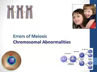
ANEUPLOID ABBERATIONS • Organism is that, which bears an irregular number of a particular chromosomes ( addition and deletion of whole sets of chromosomes). • Usually caused by failure of chromosomes to separate during meiosis (non-disjunction). • Abnormal male (XO) and female(XXY).
Non-disjunction • When a member of synaped homologous pair of chromosomes, at anaphase, fail to separate and the gametes thus formed become abnormal. • Some gametes receives both members of homologues while other gamete none. • Fertilization of such abnormal gametes from zygotes that either have an additional chromosomes(2n+1) or lack chrmosomes(2n-1).
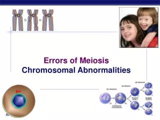
Origin of non-disjunction • An error in meiosis I leads to the gamete containing both homologs of one chromosomes pair. • In meiosis II results in the gamete receiving two copies of one of the homologs of the chromosomes pair. • Can also occur during an early mitotic division in the developing zygote, which results in mosaicism
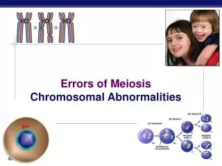
Causes of Non-disjunction • An aging effect on the primary oocyte, which can remain in a state of suspended inactivity for upto 50 years. • Association b/w advancing maternal age and increased incidence of down Syndrome.
Factors causing Non-disjunction • An absence of recombination b/w homologous chromosomes in foetal ovary • An abnormality in spindle formation • Radiation • Delayed fertilization after ovulation.
Monosomy • The absence of a single chromosome is refered to as monosomy. • Diploid organisms that has one chromosomes less than its normal diploid number(2n-1= AABBC). • Monosomies on meiosis produces two types of gametes with (n) and (n-1) chromosomes. • In animals , loss of one chromosomes often results in genetic imbalance which is associated with high mortality or reduce fertility. • Human Syndromic disease like Turners syndrome (XO) is an example of monosomic mutations. • Results due to non-disjunction in meiosis
Monosomy • If one gamete receive two copies of a homologous chromosomes, (Disomy) • While other corresponding daughter gamete will have no copy of the same chromosome (nullisomy) • Also due to loss of a chromosomes as it moves to the pole of the cell during anaphase, known as Anaphase lag.
Trisomy Presence of an extra chromosome is refered to as trisomy. • Down Syndrome (trisomy 21) • Patau Syndrome (trisomy 13) • Edward Syndrome (trisomy 18)
Trisomy • Caused by Failure of separation of one of the pairs of homologous chromosomes during anaphase of meiosis I. • Can be caused by non-disjunction occurring during meiosis II when a pair of a sister chromatids fails to separate.
Trisomy • Diploid organisms that have one chromosome extra (2n+1= AABBCCC) are called Trisomics.
Tetrasomics • Diploid organisms that have one chromosome in quarduplicate (2n+ 2= AABBCCCC).
Nullisomics • An diploid organism, that has lost one chromosome pair from its genotype is called nullisomic. • It is lethal in diploids • Some polyploids, however, can lose one homologous pair without serious effects (AAAABB) • Nullisomics of hexaploid wheat(6n-2) show reduced vigor and fertility but can survive to maturity.
Double Trisomics • If in a diploid organism, two different chromosomes are present in triplicate , called double trisomic and presented as (2n+1+1) AABBCCC • In humans Klinefelter syndrome (XXYY)
Chromosome Morphology Under the microscope chromosomes appear as thin, thread-like structures. • They all have a short arm and long arm separated by a primary constriction called the centromere. The short arm is designated as p and the long arm as q. • The centromere is the location of spindle attachment and is an integral part of the chromosome. It is essential for the normal movement and segregation of chromosomes during cell division. Human metaphase chromosomes can be categorized according to the length of the short and long arms and also the centromere location. • Metacentricchromosomes have short and long arms of roughly equal length with the centromere in the middle. • Submetacentricchromosomes have short and long arms of unequal length with the centromere more towards one end. • Acrocentricchromosomes have a centromere very near to one end and have very small short arms. They frequently have secondary constrictions on the short arms that connect very small pieces of DNA, called stalks and satellites, to the centromere. • The stalks contain genes which code for ribosomal RNA. • The diagrams showing region on chromosomes, called ideograms.
Acrocentric(Chromosome 14) Submetacentric(Chromosome 9) Metacentric(Chromosome 1)
The ideogram is basically a "chromosome map" showing the relationship between the short and long arms, centromere (cen), and in the case of acrocentric chromosomes the stalks (st) and satellites (sa). Each band is numbered to aid in describing rearrangements.
Chromosomes are identified by their size, centromere position and banding pattern Autosomes are numbered from largest to smallest, except that chromosome 21 is smaller than chromosome 22.
Cytogenetics • Is the study of the structure and properties of chromosomes, chromosomal behaviour during mitosis and meiosis, chromosomal influence on the phenotype and the factors that cause chromosomal changes. • Related to disease status caused by abnormal chromosome number and/or structure.
Methods for chromosomal analysis: Karyotyping and banding The collection of all the chromosomes is referred to as a Karyotype. The method used to analyze the chromosome constitution of an individual, known as chromosome banding. Chromosomes are displayed as a karyogram.
Obtaining and preparing cells for chromosome analysis • Cell source: • Blood cells • Skin fibroblasts • Amniotic cells / chorionic villi • Increasing the mitotic index - proportion of cells in mitosis using colcemid • Synchronizing cells to analyze prometaphase chromosomes
Key procedure • In the case of peripheral (venous) blood • A sample is added to a small volume of nutrient medium containing phytoheamagglutinin, which stimulates T lymphocytes to divide. • The cells are cultured under sterile conditions at 37C for about 3 days, during which they divide, and colchicineis then added to each culture. • This drug has the extremely useful property of preventing formation of the spindle, thereby arresting cell division during metaphase, the time when the chromosomes are maximally condensed and therefore most visible. • Hypotonic saline is then added, which causes the red blood cells to lyze and results in spreading of the chromosomes, which are then fixed , mounted on a slide and stained ready for analysis
Karyotype Analysis • Following Steps are involved; • Counting the number of cells, sometimes referred as metaphase spread • Analysis of the banding pattern of each individual chromosome in selected cells. • Total chr. Count is determined in 10-15 cells, but if mosaicism is suspected then 30 or more cell count will be undertaken. • Detailed analysis of the banding pattern of the individual chromosomes is carried out in approx. 3-5 metaphase spread, which shows high quality banding. • The banding pattern of each chromosome is specific and shown in the form of Idiogram.
Chromosome Banding • Chromosome banding is developed based on the presence of heterochromatin and euchromatin. • Heterochromatin is darkly stained whereas euchromatin is lightly stained during chromosome staining. • Euchromatin, which undergoes the normal process of condensation and decondensation in the cell cycle, and • Heterochromatin, which remains in a highly condensed state throughout the cell cycle, even during interphase.